What is Craniosynostosis?
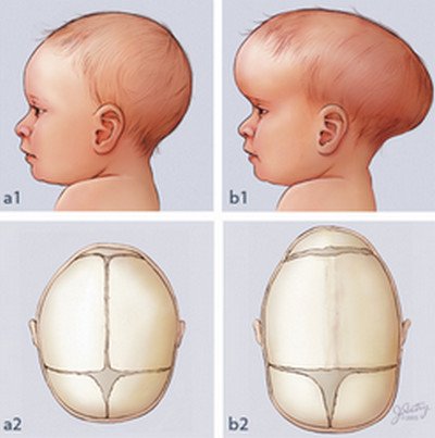
Craniosynostosis is an abnormal birth defect that causes the premature fusion of one or more sutures in a baby’s skull by ossification. This results in a distorted growth of the skull and an increase in the intracranial pressure. The fusion of the sutures would also cause the skull to develop parallel to the closed sutures instead of perpendicularly. This skull growth would give the appearance of an abnormal head shape and facial features.
Craniosynostosis Types
Craniosynostosis is classified depending on the number of fusion of sutures involved.
- Simple craniosynostosis refers to the involvement of a single suture only.
- Complex or compound craniosynostosis refers to the premature fusion of more than one suture.
- In addition, when craniosynostosis comes with deformities in other areas of the body like the limbs, heart, and central nervous system, it is termed as syndromic craniosynostosis.
- Absence of other abnormalities would be referred to as non-syndromic or isolated craniosynostosis.
Causes of Craniosynostosis
The cause of craniosynostosis is still unknown. However, several researches have suggested that genetic mutation may play a role in the development of the condition. Several factors have been identified that poses a great risk for developing craniosynostosis. These factors usually occur during pregnancy. Some of these include womb constraint, maternal smoking and exposure to drugs containing amine. Increased levels of thyroid hormones have also been associated with craniosynostosis.
Craniosynostosis Diagnosis
Craniosynostosis is diagnosed through physical assessment, medical history and radiographic examination.
- Physical assessment would show apparent signs and symptoms for craniosynostosis while medical history would check on the pregnancy condition of the mother as well as possible risk factors identified in both parents.
- Radiographic analysis of the skull can be done through computed axial tomographic scan (CT scan) although plain radiographic examination may also be utilized. The main advantages, however, of a CT scan are the easy identification of the suture and structural abnormalities of the brain.
Signs and Symptoms of Craniosynostosis
The presenting signs and symptoms of craniosynostosis are usually dependent on the type of craniosynostosis a child has.
- Generally, patients with craniosynostosis would present signs and symptoms of increased intracranial pressure (ICP), obstructive sleep apnea, skull deformities and neurobehavioral impairment.
- Vomiting, vision problems possibly leading to vision loss and headache are usually the most common symptoms for elevated intracranial pressure. The increase in intracranial pressure is usually due to the continued brain growth in a rigid skull.
- Abnormalities in the skull would include the absence of a fontanel and deformities in the shape of the head with no or slow physical growth. Physical assessment through measurement of head circumference is therefore important especially in cases with hydrocephalus and microcephaly. Physical deformities of the digits, neck, spine and toes may also be apparent especially among syndromal craniosynostosis.
- Neurobehavioral impairment would include problems with attention, visual spatial skills, reading and language. Others may present with a decreased IQ but there are also other cases in which the child presents with normal intelligence.
Craniosynostosis Pictures
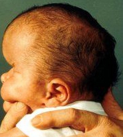
Picture 1 : craniosynostosis of the lambdoid suture
Image source: wikipedia.org
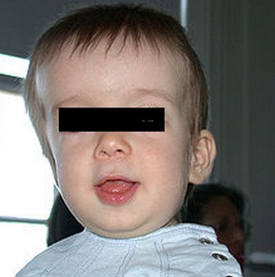
Picture 2 : Craniosynostosis
Picture 3 : Craniosynostosis image
Treatment for Craniosynostosis
Surgical intervention is the best treatment modality for children with craniosynostosis. The goals for surgery is to relieve pressure on the brain, allow space for the brain’s growth and development as well as correct and improve the physical appearance of the child. Surgical intervention is usually done when the child is still an infant and could be a series of surgeries rather than just one surgical operation.
There are various conditions which need to be met prior to surgery.
- The ideal age for surgery is between 3 to 6 months as surgery performed younger than 6 months are at an increased risk for relatively large losses of blood volume.
- The presentation of how the sutures fused prematurely also plays an important factor on when to perform the surgery.
As craniosynostosis is a life threatening condition, it is best advised that a pediatric neurologist would evaluate the condition. Monitoring of the child’s condition should start from birth onwards until such time that surgical correction is performed. In addition, craniosynostosis would require a team of specialists to monitor and evaluate the child’s condition from physical health to the mental and behavioral health of the child.
A psychologist may be necessary as the child may experience feelings of low self-esteem and depression. Regular follow up consultation is also necessary to ensure a good outcome and prognosis for the child.
References:
http://www.ncbi.nlm.nih.gov/pubmedhealth/PMH0002557/
http://emedicine.medscape.com/article/1175957-treatment#showall