Introduction
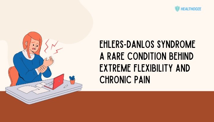
Ehlers-Danlos Syndrome (EDS) is a group of rare inherited conditions that affect connective tissue strength and elasticity. Connective tissues give structure and support to the body’s organs, joints, and skin.
People with EDS often have highly flexible (“double-jointed”) limbs, fragile skin, and persistent discomfort. Although the word “flexibility” might sound positive, the extreme mobility in EDS can lead to frequent dislocations, muscle strains, and sometimes significant pain.
EDS is not always recognized immediately. Symptoms may appear mild in childhood but become more noticeable with daily activities or minor injuries. Individuals might endure joint instability, prolonged wound healing, or fatigue without knowing a genetic cause exists.
This lack of awareness can delay diagnosis, reduce access to proper treatments, and cause emotional stress. In recent years, better understanding of connective tissue disorders has shed light on EDS, improving clinical guidelines and management strategies.
This article provides an overview of EDS, explains its genetic background, and outlines how patients and their families can navigate the physical and emotional challenges. By learning about EDS, readers may better identify and support people living with this rarely understood condition.
What Is Ehlers-Danlos Syndrome?
Ehlers-Danlos Syndrome (EDS) is an inherited group of disorders that compromise the structure or function of collagen, a key protein in connective tissue. Collagen acts like the “glue” that holds tissues together. When it is faulty or underproduced, tissues become too elastic or fragile. Different EDS types exist, each varying in specific collagen-related gene mutations. However, they share a general tendency for overly flexible joints, loose or delicate skin, and a range of possible internal complications.
Historically, EDS has been misdiagnosed or overlooked because its signs can resemble more common orthopedic or dermatological problems. Joint dislocations, for instance, might be attributed to sports injuries, while easy bruising could be mistaken for a clotting disorder. In many cases, distinct patterns—like unusual flexibility, frequent subluxations, or an apparent family history—signal a connective tissue problem that warrants thorough genetic and clinical evaluation.
Although EDS is considered “rare,” some forms, such as hypermobile EDS, may be more prevalent than once thought. Emerging data suggest that a spectrum of hypermobility disorders overlaps with EDS features. For example, some people have significant joint laxity and mild symptoms that do not fit precisely into classic categories. These borderline presentations further complicate EDS awareness and diagnosis.
EDS is not solely about joints or skin. Connective tissue permeates the entire body, so patients can experience an array of complications. These might involve blood vessel walls, gastrointestinal linings, or uterine tissues in women. Understanding this complexity helps with anticipating potential health risks and planning preventive measures over a lifetime.
Types of Ehlers-Danlos Syndrome
EDS encompasses more than a dozen classifications, though certain types occur more frequently than others. The current categories, defined by clinical criteria and genetic findings, include:
- Hypermobile EDS (hEDS)
- Most common type
- Dominant features: joint hypermobility, subluxations, and varying degrees of pain
- Skin may be only slightly affected, leading to fewer external markers
- Classical EDS (cEDS)
- Characterized by highly elastic, soft, and often translucent skin
- Wounds may heal slowly, leaving wide or atrophic scars
- Joint hypermobility typically affects both large and small joints
- Vascular EDS (vEDS)
- Notable for fragile blood vessels and organ membranes
- High risk of arterial or organ rupture, making it potentially life-threatening
- Relatively rare but demands specific caution regarding physical activity
- Kyphoscoliotic EDS (kEDS)
- Prominent spinal curvature (kyphoscoliosis) visible early in life
- Joint laxity and muscle weakness
- Potential for severe mobility limitations over time
- Arthrochalasia EDS (aEDS)
- Extreme hypermobility affecting multiple joints, especially the hips
- Early onset of bilateral hip dislocations
- Skin hyperextensibility may be milder than in other types
- Dermatosparaxis EDS (dEDS)
- Noticeable skin fragility, with sagging and soft texture
- High risk of hernias and large scars
- Collagen processing defect leads to severe skin manifestations
- Other Rare Types
- Some EDS variants are extremely unusual, combining distinctive features like gum fragility, short stature, or severe progressive scoliosis.
- Genetic testing can help distinguish these unique subtypes.
Each type follows its own pattern of inheritance—most often autosomal dominant, though recessive inheritance appears in certain subtypes. Precise genetic characterization continues evolving, with newer EDS-related mutations discovered in research. Genetic counseling offers families a clearer perspective on recurrence risks and the variability among their relatives.
Causes and Genetics
At the root of Ehlers-Danlos Syndrome are genetic defects affecting collagen synthesis, structure, or processing. Collagen is one of the most abundant proteins in the body, helping maintain form and stability in skin, ligaments, tendons, and blood vessel walls. When collagen is faulty, the tissue’s normal elasticity is disturbed, leading to hypermobility or fragility. Some EDS subtypes involve defects in enzymes that modify collagen after it is produced, further disrupting the tissue’s strength.
Collagen Basics
- Structural Protein: Collagen provides a supportive framework for cells and tissues.
- Triple Helix Formation: Typical collagen molecules form a rope-like triple helix, imparting strength.
- Mutation Effects: A single amino acid substitution or a missing enzyme can unravel the triple helix or reduce collagen quantity.
Inheritance Patterns
- Autosomal Dominant: A single defective gene from one parent can cause the condition. Examples: classical EDS, hypermobile EDS.
- Autosomal Recessive: Two copies of the defective gene (one from each parent) must be inherited. Examples: dermatosparaxis EDS, some forms of kyphoscoliotic EDS.
In certain families, the condition surfaces each generation, indicating a dominant pattern. However, some children with unaffected parents might have a de novo mutation—an alteration in the gene that arises spontaneously in the egg, sperm, or early embryo. Genetic testing, while helpful, is not yet comprehensive for all EDS types. Hypermobile EDS remains tricky, as no single gene test is consistently definitive.
Environmental factors do not directly cause EDS, but they may influence symptom severity. For instance, repeated injuries can worsen joint instability, or poor nutritional support might accelerate tissue breakdown. Hormonal fluctuations may also aggravate joint laxity, especially in women during pregnancy or postpartum.
Signs and Symptoms: Beyond Joint Hyperextensibility
Although the hallmark of EDS is unusual joint mobility, the condition extends to multiple body systems. Recognizing the variety of symptoms can lead to earlier suspicion and a thorough examination.
Musculoskeletal Manifestations
- Chronic Joint Pain
- Dull aches or sharp pains in knees, hips, shoulders, or small joints
- Often worsens with prolonged standing or repetitive motions
- Frequent Dislocations or Subluxations
- Simple movements can cause the joint to slip out of place
- Shoulders, knees, ankles, and fingers frequently affected
- Muscle Fatigue
- The body expends extra energy stabilizing lax joints, leading to exhaustion
- Soft tissue injuries or tendonitis might appear after mild activity
Dermatological Features
- Soft, Velvety Skin
- In classical types, the skin may have a plush feel
- Skin is easily indented or pinched beyond normal range
- Delayed Wound Healing
- Scar tissues tend to be wide or “cigarette paper” in appearance
- Minor cuts can take longer to close, risking infection or overgrown scars
- Bruising and Easy Marking
- Fragile tissues tear or bruise with minimal trauma
- Subcutaneous bleeding might occur spontaneously in vascular EDS
Cardiovascular and Organ Involvement
- Arterial Fragility
- In vascular EDS, arteries may develop aneurysms or experience spontaneous rupture
- Arterial dissection can emerge at younger ages than expected
- Gastrointestinal Complications
- Bowel or stomach tissue fragility can lead to chronic digestive issues, hernias, or organ prolapse
- Recurrent abdominal pain or severe constipation may indicate structural problems
- Pelvic and Uterine Issues
- Women may notice uterine prolapse, vaginal tears during childbirth, or postpartum hemorrhage
- Hormonal changes and pregnancy place additional stress on connective tissues
Neurological and Autonomic Effects
- Headaches
- Tension headaches or migraines might be worsened by joint instability in the cervical spine
- Craniocervical instability in certain EDS forms can compress nerve structures
- Autonomic Dysfunction
- Dysregulated blood pressure, heart rate, or temperature regulation
- Possible link with postural orthostatic tachycardia syndrome (POTS)
- Sensory Oversensitivity
- Heightened pain perception or tactile sensitivity
- Some individuals experience heightened responses to certain fabrics or minor touch
The interplay of these symptoms can be debilitating and complex. Over time, repeated injuries and chronic pain accumulate, diminishing the individual’s capacity for routine tasks. Pain management, physiotherapy, and lifestyle adaptations become crucial for preserving function and reducing the risk of further complications.
Diagnosis: A Complex Journey
EDS can be elusive. Doctors may focus on a patient’s single complaint—like knee pain—without recognizing an underlying connective tissue disorder. Individuals, too, might not suspect a unifying genetic cause if the family history is incomplete or if mild forms run in older relatives who never sought diagnosis.
Clinical Assessment
- Physical Examination
- Joint mobility measured using tools like the Beighton Score (which checks flexibility in joints such as the thumb, elbows, knees)
- Skin extensibility tested gently, often comparing to typical ranges
- Medical History
- Reviewing patterns of past injuries, surgeries, or scarring
- Learning about family members with unusual joint problems, heart complications, or fragile skin
- Systemic Evaluation
- Checking for signs of organ fragility, vascular anomalies, or neurological issues
- Ruling out other conditions with overlapping features (e.g., Marfan syndrome, osteogenesis imperfecta)
Genetic Testing
While genetic tests are definitive in some EDS subtypes (e.g., vascular, kyphoscoliotic), hypermobile EDS often lacks a single confirmatory test. Even if a gene panel is negative, a clinical diagnosis might still be made based on recognized patterns and physical findings.
The Role of Specialists
A multidisciplinary team is often required:
- Rheumatologist: Evaluates connective tissue and autoimmune factors
- Geneticist: Interprets test results and counsels on inheritance patterns
- Cardiologist: Screens for vascular complications, especially in suspected vEDS
- Orthopedist: Assesses chronic joint instability and potential surgeries
Recognition by healthcare professionals that EDS is a valid, multi-system disorder speeds up accurate diagnosis and better outcomes. However, late diagnoses remain common. Many patients express relief when they finally name the cause behind years of unexplained injuries and pain.
Managing EDS: Strategies and Approaches
There is no cure for Ehlers-Danlos Syndrome. Treatment aims to protect joints, handle pain, and address complications preemptively. Personalizing care helps individuals maintain the best possible quality of life.
Physical and Occupational Therapy
- Physiotherapy
- Gentle muscle-strengthening exercises support unstable joints
- Targeted routines focus on the core, hips, and shoulders to stabilize frequently subluxed areas
- Occupational Therapy
- Teaches adaptive methods for daily activities, reducing strain on fragile tissues
- Might suggest specialized devices (e.g., jar openers, supportive braces)
- Joint Protection Techniques
- Avoiding overstretching or forceful movements that risk dislocation
- Guidance on lifting objects safely with minimal stress on wrists and elbows
Pain Management
- Medication
- Over-the-counter pain relievers (e.g., acetaminophen)
- Prescribed nonsteroidal anti-inflammatory drugs (NSAIDs)
- Opioids used cautiously and under close supervision
- Some patients find relief with nerve-targeting medications, like certain antidepressants or anticonvulsants
- Topical Treatments
- Creams or patches containing analgesic ingredients can soothe localized muscle aches
- Heat or cold therapy to calm inflamed areas
- Alternative Therapies
- Acupuncture or massage: may temporarily ease pain, though results vary
- Mind-body practices (yoga, tai chi) performed carefully under trained instructors
Lifestyle Modifications
- Exercise and Activity Balance
- Low-impact aerobic exercise (swimming, stationary cycling) builds endurance without excessive stress on joints
- Gradual increments in intensity to prevent flare-ups
- Rest and Recovery
- Scheduling breaks or rests between tasks
- Listening to early pain signals and pausing activity can avert worse damage
- Bracing or Splints
- Stabilizes problematic joints (knees, wrists, ankles)
- Helps limit subluxations during activities like typing, cooking, or sports
- Environmental Adjustments
- Ergonomic furniture and supportive mattresses
- Minimizing tripping hazards or environmental factors that heighten risk of falling or collisions
Surgical Interventions
- Joint Stabilization: In recurrent dislocations, surgical tightening of tendons or ligaments might be considered
- Vascular Repairs: In vEDS, aneurysms or arterial tears may need urgent vascular surgery
- Caution: Surgery carries extra risks in EDS (e.g., slow healing, scar fragility), so thorough planning with EDS-knowledgeable specialists is essential
Emotional and Social Implications
Living with EDS can take a toll on emotional well-being. Chronic pain, fatigue, and repeated injuries disrupt normal routines, diminishing a person’s sense of independence or identity. Younger patients may struggle to keep up with peers in sports or daily tasks, feeling singled out or frustrated.
Mental Health Concerns
- Anxiety
- Fear of joints dislocating or the unpredictability of vascular issues
- Worry about needing urgent medical care without a moment’s notice
- Depression
- Persistent pain and physical limitations can lead to low mood
- Social isolation if pain or fatigue curtails outings, jobs, or school attendance
- Body Image
- Scarring or fragile skin affects self-esteem
- Using braces or mobility aids can lead to self-consciousness, especially in teenage or early adult years
Coping Strategies
- Counseling and Therapy
- Psychologists experienced with chronic health conditions can help patients develop resilience
- Cognitive-behavioral therapy (CBT) addresses negative thinking patterns and fosters better coping
- Support Groups
- Online and in-person communities connect those with EDS for shared resources, tips, and emotional backing
- Learning from others reduces feelings of isolation
- Educational Accommodation
- Younger patients might need flexible schooling or an individualized plan due to pain or mobility constraints
- Tools like dictation software reduce strain on wrists or fingers
- Workplace Adjustments
- Modified schedules, telecommuting, or desk setups that reduce repetitive stress
- Communicating with employers about occasional flare-ups or doctor’s appointments
Life with EDS: Practical Tips
Every individual’s experience differs. However, some general methods help manage day-to-day life with Ehlers-Danlos Syndrome:
- Know Your Limits: Overexertion can worsen symptoms. Learn early warning signs (like muscle tension or joint instability) that suggest you need rest.
- Stay Hydrated and Maintain Nutrition: Water intake can help with blood pressure issues (if there’s autonomic involvement). A balanced diet supports tissue repair.
- Plan for the Long Term: If traveling or performing physically intense tasks, schedule more breaks and research medical facilities in advance.
- Seek Expert Opinions: Connect with doctors knowledgeable in EDS or consult EDS-specific clinics. They may offer advanced imaging, bracing solutions, or targeted therapies.
- Build a Reliable Health Team: Physiotherapists, mental health professionals, orthopedists, and sometimes a pain management specialist. Frequent communication fosters a holistic approach.
- Emphasize Self-Care: Gentle stretches, warm baths, or mindfulness exercises can calm stress and ease muscle tension.
- Stay Updated on Research: EDS is an evolving field. Periodically check reputable sources or patient networks to learn about new treatments or guidelines.
Table: Key EDS Types and Main Features
| Type | Key Characteristics | Inheritance Pattern |
| Hypermobile (hEDS) | Joint hypermobility, subluxations, moderate skin involvement, no single definitive gene test | Usually autosomal dominant |
| Classical (cEDS) | Highly elastic skin, widespread atrophic scarring, joint hypermobility | Autosomal dominant |
| Vascular (vEDS) | Fragile arteries/organs, risk of rupture, thin translucent skin | Autosomal dominant |
| Kyphoscoliotic (kEDS) | Congenital muscle weakness, scoliosis, joint laxity, potential eye involvement | Autosomal recessive |
| Arthrochalasia (aEDS) | Severe hypermobility, early bilateral hip dislocations, mild skin involvement | Autosomal dominant |
| Dermatosparaxis (dEDS) | Very fragile skin with sagging, delayed wound healing, hernias | Autosomal recessive |
Conclusion
Ehlers-Danlos Syndrome is far more than “bendy joints.” It is a spectrum of genetic connective tissue disorders that impact daily life through chronic pain, dislocations, delicate skin, and potential organ risks. The broad range of symptoms calls for a team-based approach, integrating physical therapies, pain management, lifestyle modifications, and emotional support. Timely diagnosis can prevent repetitive injuries and guide more effective care, yet individuals often face confusion or misdiagnosis due to the condition’s rarity and variability.
Increasing awareness is key. Greater clinician education about EDS fosters quicker recognition, while family and peer understanding reduces stigma. Although living with EDS presents ongoing challenges—from possible surgeries to coping with chronic discomfort—many adapt their environments, set realistic goals, and maintain fulfilling lives. Ongoing scientific research offers optimism that improved genetic insights and new therapeutic methods will further enhance outcomes for people with EDS. By embracing empathy and evidence-based care, we can support and empower individuals who navigate this rare yet significant disorder.
References
- Beighton P, De Paepe A, Steinmann B, Tsipouras P, Wenstrup RJ. Ehlers-Danlos syndromes: Revised nosology, Villefranche, 1997. Am J Med Genet. 1998;77(1):31–37.
- Malfait F, Francomano C, Byers P, et al. The 2017 international classification of the Ehlers–Danlos syndromes. Am J Med Genet C Semin Med Genet. 2017;175(1):8–26.
- Castori M, Morlino S. Ehlers-Danlos syndrome hypermobile type: a comprehensive framework for clinical diagnosis and management. Am J Med Genet C Semin Med Genet. 2019;181(1):4–13.
- Colombi M, Dordoni C, Chiarelli N, et al. Aortic aneurysm in classic Ehlers-Danlos syndrome: report on a family harboring a COL5A2 mutation. Hum Mutat. 2017;38(2):147–151.
- De Paepe A, Malfait F. The Ehlers-Danlos syndrome, a disorder with many faces. Clin Genet. 2012;82(1):1–11.
- Norris RA, Smolarek T, Brodsky GL, et al. Ehlers–Danlos syndrome and related connective tissue diseases: understanding collagen gene structure and function. J Med Genet. 2014;51(3):90–103.
- Tinkle B, Berglund B, Francomano C, et al. Hypermobile Ehlers-Danlos syndrome (a.k.a. Ehlers-Danlos syndrome Type III and Ehlers-Danlos syndrome hypermobility type): Clinical description and natural history. Am J Med Genet C Semin Med Genet. 2017;175(1):48–69.
- Hakim AJ, Grahame R. A simple questionnaire to detect hypermobility: an adjunct to the assesment of patients with diffuse musculoskeletal pain. Int J Clin Pract. 2003;57(3):163–166.
- Tinkle BT, Levy HP. Symptomatic joint hypermobility: the hypermobile type of Ehlers–Danlos syndrome and the hypermobility spectrum disorders. Med Clin North Am. 2019;103(6):1021–1033.
- Chopra P, Tinkle B, Hamonet C, et al. Pain management in the Ehlers–Danlos syndromes. Am J Med Genet C Semin Med Genet. 2017;175(1):212–219.
- Bloom L, Byers PH. Vascular Ehlers-Danlos syndrome. GeneReviews. 2018.
- Berglund B, Bonk K, Domingues DF, et al. Physiotherapy management for hypermobility spectrum disorders including hypermobile Ehlers-Danlos syndrome. Am J Med Genet C Semin Med Genet. 2021;187(4):433–447.

