
What is Erythroplakia?
Erythroplakia refers to an oral condition that appears as red lesions on the mucus membrane but cannot be attributed to any other illness or condition [1].
The reddish color of the patches has been attributed to lack of a keratin layer on the mucosal tissue. Instead of the keratin, there is a papillae connective tissue with enlarged capillaries. The capillaries happen to be in close proximity to the surface and that also plays a role in the reddish color [2].
The condition is similar to leukoplakia, which is characterized by white spots on the mucous membrane of the mouth [1, 3].
The condition is prevalent among men between the age of 60 and 70 years [2]. Together with leukoplakia, erythroplakia is considered to be a premalignant lesion [4]. As such, timely diagnosis and treatment is of great important to avoid development of cancer in the lesions. Routine oral check-ups will ensure the condition is detected early enough [5].
What does Erythroplakia look like (Pictures) ?
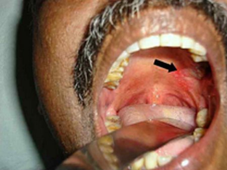
Image 1: Erythroplakia lesions on the palate (Indicated by the arrow)
Photo Source: screening.iarc.fr

Image 2: Erythroplakia on the tongue
Picture Source: screening.iarc.fr
History of Erythroplakia
- A non-hemorrhagic, painless, red color lesion was observed on the upper part of the gut. This was observed and reported in 1852 [2].
- Nevertheless, the first medical description of the condition in an academic paper was done in the year 1911 by Queyrat. In the paper, Queryat described a red patch on the glans of the penis of a person with syphilis. Queyrat coined the term erythroplakia [6].
- Rubin named incipient carcinoma with the aim of describing the erythroplakia microscopic properties he observed in the same era. The precancerous nature of the lesions has become the most widely used histo-pathologic diagnosis Erythroplakia [2].
Sings and Symptoms
- Erythroplakia is asymptomatic [4].
Signs
- Red patches on the mucosal and/or sub-mucosal surfaces
- Occurrence of pain may be an indication that the lesions are entering the malignant phase [2].
Cause:
- It is idiopathic thus may come up spontaneously without a known cause [2, 6].
Risk Factors include
- Smoking tobacco
- Excessive consumption of alcohol
- An opportunistic infection or a candidiasis super-infection. This could be associated with the oral-mucosal surface cells. Candida albicans has been observed severally in the erythroleukoplakia patches. The white and/or the red components of the lesions may diminish after the patient has received antifungal therapy [3, 7].
Clinical Features of Erythroplakia
1. Occurrence varies with:
- Age: mean age at diagnosis of erythroplakia is 50-69 years, with less than 5% of diagnoses patients being under 30 years of age. Older patients also have a higher the risk of for the lesions to undergo the premalignant to malignant transformation.
- Sex: Erythroplakia is predominant among men. However, recent ratios show an increase in female patients, possibly owing to an increase in female smokers [8].
2. Sites:
- Generally, Erythroplakia can occur on any of the mucosal surfaces around the neck or head [2].
- However, up to half of the diagnosed cases have been noted to exist on vermilion and intraoral surfaces. The rest of the recorded occurrences are divided between larynx and the pharynx.
- Vermillion patches are quite common and are most often found on the lower lips.
3. Homogenous form:
- Usually found on the buccal mucosa and the soft palate. Sometimes they are found on the tongue or the floor of the mouth, albeit rarely. [2].
- This form is occurs as a soft, red, and velvety lesion with straight margins that are well-demarcated.
- Can be quite large in size in terms of the area they cover.
4. Size:
Usually less than 15mm in diameter. However, some lesions are more than 4cm in diameters.
5. Margins:
The lesions contrast sharply with the normal pink mucosa surroundings in the mouth. [3, 7].
6. Granular/Speckled form:
They comprise of red and soft lesions. They are raised slightly and have regular outlines. The surface is finely nodular and speckled tiny plaques which are white in color.
7. Smooth erythroplakia:
A palpitation test reveals a soft texture which is commonly described as a velvety feel on the fingers. The pebbled patches/lesions are a little firm but the erythroplakia doesn’t become really indurated until an invasive carcinoma develops within [2].
8. Erythroleukoplaka:
- This condition is commonly observed where erythroplakia appears adjacent to or in same area as a leukoplakia in the mouth [9].
- The red spots are most probable to contain or develop dysplastic cells. Therefore, the red areas have to be biopsied readily and clinically examined with uttermost care.
- Erythroleukoplaka patches are quite irregular and usually not as bright as the erythroplakia homogenous type. They are found on the floor part of the mouth and on the tongue in most occasions.
- The borders are not as clear as in the homogenous lesions and they may blend with the surrounding oral mucosa surface.
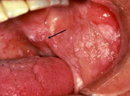
Image 3: Erythroleukoplakia
Photo Source: medscape.com
9. Unlike the leukoplakia lesions, erythroplakia covers large areas in the mouth. In addition, it rarely expands laterally after the initial diagnosis. However, it might be an artifactual property because most of the lesions are destroyed or removed totally after diagnosis.
Diagnosis
- Erythroplakia rarely covers large parts of the patient’s mouth. While examined by use of hand, the erythroplakia feels soft. It remains soft without hardening till a persistent carcinoma develops inside the lesion [10].
- Although some of the people with the condition may experience a burning, or sore sensation, erythroplakia is asymptomatic. Oral erythroplakia possesses the greatest risk of attaining malignancy compared to the other types of premalignant lesions.
- Medical examination of the diagnosed erythroplakia reveals severe dysplasia of the epithelium or invasive cancer [11]. All erythroplakia lesions should be examined for malignancy with extreme clinical caution and suspicion. This is because erythroplakia lesions are highly likely to contain the histological foci of chronic dysplasia, or even invasive cancer.
- Excision biopsy is advised and proper histological examination should follow as a compulsory practice every time there is clinical diagnosis of erythroplakia.
Treatment and Management
- Treatment involves biopsy of the lesion to identify extent of dysplasia [10].
- Complete excision of the lesion is sometimes advised depending on the histopathology found in the biopsy. Even in these cases, recurrence of the erythroplakia is common and, thus, long-term monitoring is needed.
- The management of the condition involves taking measures that avoid the transformation into malignancy [7]. Creating public awareness about the illness, closely monitoring those that have undergone treatment and regular oral check-ups are sure ways of controlling it [4, 8].
References:
- http://www.oralcancerfoundation.org/cdc/cdc_chapter4.php
- http://dentalproblems.ygoy.com/2011/10/18/oral-precancer-erythroplakia-overview
- http://emedicine.medscape.com/article/1840467
- http://www.ada.org.au/app_cmslib/media/lib/1108/m324364_v1_villa.pdf
- http://www.webmd.boots.com/cancer/cancer-of-the-mouth-and-throat-overview
- https://en.wikipedia.org/wiki/Erythroplakia
- http://screening.iarc.fr/atlasoral_list.php?cat=A3&lang=1
- http://www.cancer.ca/en/cancer-information/cancer-type/oral/oral-cancer/precancerous-conditions/?region=on
- http://www.kcl.ac.uk/dentistry/about/acad/oral-leukoplakia-and-erythroplakia.pdf
- http://www.rightdiagnosis.com/e/erythroplakia
- http://www.healthcommunities.com/head-and-neck-cancer