The heart is one of the vital organs in the human body. It pumps blood throughout the body and supplies nutrients and oxygen to the tissues. Oxygenated blood is needed by the body to be active and perform its vital functions. The heart beats about 72 times per minute.
The heart is a muscle that expands and contracts. The pumping of the heart is referred to as the cardiac cycle and each cycle lasts eight-tenths of a second. (1, 2, 3)

Image 1: The clinical presentation of the human heart.
Picture Source: img.purch.com

Picture 2: The different parts of the human heart.
Photo Source: www.worldinvisible.com

Photo 3: The human heart highlighting the four heart valves.
Image Source: www.cardiosmart.org
The Anatomy of the heart
- Measurement – The size of the heart is the same as the size of the large fist. It weighs between 10 to 12 ounces in men and 8 to 10 ounces in women.
Chambers – There are four chambers in the heart; two atria (upper chamber) and two ventricles (lower chambers).
- Right atrium and right ventricle – consist the right side of the heart.
- Left atrium and left ventricle – consist the left side of the heart.
- Septum – it is a wall muscle that separates one side of the heart to the other.
- Pericardium – it encases, protect, and anchor the heart inside the chest.
- Pericardial fluid – it lubricates the heart during contractions.
- Outer wall – The outer wall of the heart has three layers:
- Epicardium – the outermost wall layer.
- Myocardium – the middle layer containing the muscle used for contraction.
- Endocardium – inner layer of the heart; the one that gets in contact with the blood. (2, 3, 4, 5)
Heart valves – The heart has four valves:
- Mitral valve and tricuspid valve (Atrioventricular valves) – They are found between the atria and the ventricles. They control the flow of blood from the atria to the ventricles.
- Aortic valve and pulmonary valve (semilunar valves) – They are found between the ventricles and the major blood vessels. They control the flow of blood out of the ventricles.
Conduction of the heart
The heart has the ability to set its own rhythm and signal needed to coordinate the rhythm.
- Sinoatrial node – It produces an electric pulse causing the heart to contract. It is the pacemaker of the heart. It is found in the wall of the right atrium just below to the superior vena cava.
- Atrioventricular node – It is located in the right atrium, just below the interatrial septum. It picks up the signal sent by the sinoatrial node and send it to the AV bundle.
- Purkinje fiber – it carries the signal to the ventricular wall and stimulates the muscles of the heart to contract in a regular way so as to effortlessly pump blood out of the heart going to the different systems of the body. (3, 4, 5, 6)

Image 4: A diagram outlining the cardiac cycle.
Picture Source: i.pinimg.com
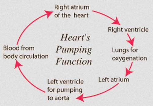
Picture 5: The pumping action of the heart.
Photo Source: hyperphysics.phy-astr.gsu.edu
The physiologic component of the heart
- Coronary systole and diastole
- Systole – it is a phase in which the muscles of the heart contract to push blood out of the chambers of the heart. During systole phase, the blood pressure rises.
- Diastole – It is a phase in which the cardiac muscles are relaxed thereby allowing the chambers of the heart to be filled with blood. During the diastole phase, the blood pressure lowers. (5, 6, 7)
The cardiac cycle
it pertains to the events that takes place during one heartbeat. The cardiac cycle has three phases:
- Atrial systole – The atria contract and push the blood into the ventricles. The AV vales are opened and the SV valves are closed to prevent the blood from re-entering the heart. The atria, being smaller than the ventricles can only fill 25% of the ventricle during the atrial systole phase.
- Ventricular systole – The ventricles contract and push blood into the aorta and pulmonary trunk. As the pressure in the ventricles increases, the semilunar valves are forced to open and the AV valves close thereby allowing the blood to flow from the ventricles into the arteries.
- Relaxation phase – In this phase, all the chambers of the heart are in the diastole enabling the blood to pour into the heart from the veins. During this phase, about 75% of the ventricles are filled with blood and will be filled completely once the atria enters systole. It is also in the relaxation phase where ventricles’ muscle cells repolarize and prepare for the next round of contraction. (7, 8, 9, 10)
The sounds of the heart
The normal heartbeat’s sound is interpreted as “lubb-dupp.” The lubb is longer than dupp and is produced as the AV valves close at the start of the ventricular systole. The dupp is the shorter and sharper sound and is produced as the semilunar valve closes at the end of the ventricular systole.
By listening to the sounds the heart produced, a cardiologist will be able to find out the presence of structural problems in the heart. (1, 4, 7)
Key points to remember:
- The size of the human heart is the same as the size of the large fist.
- The heart beats around 115,000 times per day or about 3 billion beats in a lifetime.
- The newborn’s heartbeat is faster than the adult. The adult heart beats around 60 to 80 per minute while the newborn’s heart beats around 70 to 190 beats per minute.
- The heart pumps about 5.7 liters of blood throughout the body or 2,000 gallons of blood every day.
- The heart can still beat even if it is taken out from the body.
- Heart attack is the common heart problem and it usually occurs on a Monday and usually on Christmas Day.
- The heart is situated in the center of the chest but is slightly pointing to the left.
- The woman’s heart beats faster than men.
- It is possible to die from a broken heart but is rare.
- The cells of the heart stop dividing, which makes it impossible to have a cancer of the heart.
- Laughing is a good exercise for the heart. (3, 6, 9, 10)
References:
- https://www.livescience.com/34655-human-heart.html
- https://en.wikipedia.org/wiki/Heart
- https://www.worldinvisible.com/apologet/humbody/heart.htm
- http://www.teachpe.com/anatomy-physiology/the-circulatory-system/the-heart
- https://www.acls.net/anatomy-of-the-human-heart.htm
- https://www.hopkinsmedicine.org/healthlibrary/conditions/cardiovascular_diseases/basic_anatomy_of_the_heart_85,P00192
- http://www.interactive-biology.com/75/show-me-a-diagram-of-the-human-heart-here-are-a-bunch/
- https://www.webmd.com/heart/picture-of-the-heart
- https://health.howstuffworks.com/human-body/systems/circulatory/heart.htm
- https://sciencetrends.com/parts-human-heart-vital-functions/