Archive for December, 2017

What is Transvaginal Mesh

Dec 23 2017 Published by under Women's Health

The pelvis is a delicate area because it encloses essential organs such as the bladder, vagina, and rectum. The muscles supporting the pelvic area are called pelvic floor muscles.

In some circumstances, women may experience pelvic floor dysfunction. Studies showed that about 50% of women who have undergone child birth experienced pelvic floor dysfunction or pelvic organ prolapse.

A dysfunction in the pelvic floor can also be due to hormonal changes, especially when a woman go through menopause. Performing strenuous activities such as heavy lifting for a long period of time can also affect the tightness and elasticity of the pelvic muscles. Obese women are prone to pelvic floor dysfunction. (1, 2, 3)

An image illustrating pelvic floor dysfunction Transvaginal Mesh photo picture

Photo 1: An image illustrating pelvic floor dysfunction.
Picture Source: www.holisticpaindoc.com

Pelvic floor dysfunction symptoms include the following:

  • Involuntary urine leakage with simple activities such as coughing, sneezing, or walking.
  • Pain, discomfort, and a feeling of heaviness in the vaginal area.
  • Sexual intercourse is extremely difficult for women secondary to pain and discomfort.
  • In severe cases, women have a total loss of sensation, which makes it extremely difficult to push when urinating. (2, 3, 4)

To find relief, patients should undergo reconstructive surgery. One of the commonly used ways to correct pelvic floor dysfunction is by implanting a transvaginal mesh. It is also called urogynecological mesh, pelvic mesh, and bladder sling. (3, 4)

Women who have had numerous child birth are prone to pelvic floor dysfunction Transvaginal Mesh image picture photo

Image 2: Women who have had numerous child birth are prone to pelvic floor dysfunction.
Photo Source: pbs.twimg.com

Extremely overweight-obese women are prone to pelvic floor dysfunction Transvaginal Mesh image picture photo

Picture 3: Extremely overweight/obese women are prone to pelvic floor dysfunction.
Image Source: static.dnaindia.com

Who are at risk for pelvic floor dysfunction?

  • Those who have family history of pelvic floor dysfunction.
  • Those who have undergone child birth, especially those who have multiple child birth.
  • Pelvic floor dysfunction is linked with hormonal imbalance, which is common in women in their menopausal stage.
  • Extremely overweight/obese women.
  • Those who are exposure to strenuous activities for a long period of time.
  • Women in their geriatric years. The tissues and ligaments surrounding the pelvic organs become weak with age.
  • Women who have been coughing for a long period of time such as in the case of chronic smokers. (4, 5, 6, 7)

A closer look at a transvaginal mesh image photo picture

Photo 5: A closer look at a transvaginal mesh.
Picture Source: dss.fosterwebmarketing.com

What is transvaginal mesh?

A transvaginal mesh is an implant that corrects pelvic floor dysfunction such as stress urinary incontinence and pelvic organ prolapse. It is a net-like implant available in various forms such as tape, ribbon, sling, mesh, and hammock. It is made from synthetic polypropylene. A transvaginal mesh is implanted in women to hold up a loose and weakened muscles and tissues in the pelvic area. Implanting transvaginal mesh can be done in two ways:

  • Transabdominal (abdominal route) – A small abdominal incision is created and a mesh is implanted in proper place. (5, 6)
  • Transvaginal (vaginal route) – The vagina is opened and the mesh is inserted and placed in a secure place. It helps treat urinary incontinence by creating an angle between the urethra and bladder. (6, 7)

A transvaginal mesh is used to treat and manage pelvic organ prolapse image photo picture

Picture 6: A transvaginal mesh is used to treat and manage pelvic organ prolapse.
Image Source: i.ytimg.com

What is transvaginal mesh used for?

  • Stress urinary incontinence
  • Bladder prolapse
  • Pelvic organ prolapse (6, 7, 8)

What could go wrong?

A mesh implant had been used in other parts of the body and there were no complications, adverse reactions, or injuries reported. However, the mesh reacts differently when inserted in the abdomen and in the vagina.

There are reported cases of serious physical complications in women who have undergone surgical mesh implants. Pelvic mesh symptoms include:

  • Erosion
  • Perforation of the organ in the pelvic region
  • Pain in the pelvic area
  • Vaginismus (painful sexual intercourse and trouble inserting anything in the vaginal area)
  • Inability to sit (4, 5)
  • Pudendal and obturator nerve damage
  • Neuralgia (painful nerve pathway)
  • Vaginal infection and bleeding
  • A feeling that something is protruding from the genital area
  • The pain in the vaginal area gets severe with movement and exercise (9, 10)

The two primary complications of transvaginal mesh are erosion and organ perforation. Erosion takes place when the mesh gets in contact in the patient’s soft internal tissues. The patient will complain of severe pain. The mesh protrudes through the vagina. In a vaginal examination, you will see a part of the mesh in the vaginal wall.

For the patient to experience relief from erosion, the transvaginal mesh should be removed as soon as possible before it can affect other delicate organs in the pelvic area. The problem with transvaginal mesh removal is that it is an extremely delicate procedure.

Most of the time, it will require numerous surgeries for the mesh to be completely removed. In fact, surgery does not guarantee that the mesh will be removed completely.

Organ perforation is another serious complications of transvaginal mesh. In a severe case, the mesh can cause injuries to the surrounding organs, especially the bladder, urethra, and rectum. If multiple organs are severely damaged, the patient will experience breathing problems and severe infection.

Depending on the severity of the infection, the patient might need another surgery apart from surgical removal of the mesh. Blood transfusion might be necessary too, especially if the patient has severe blood loss.

These transvaginal mesh complications can significantly affect the way patient’s way of life. Given the surgical mesh complications symptoms mentioned above, it would be extremely impossible for women to perform activities of daily living. Social and intimate relationship will be significantly affected too.

Because of these transvaginal mesh problems, the Food and Drug Administration (FDA) issued a warning against the use of transvaginal mesh. It is a high-risk device and those who are planning to have it should know the pros and cons of transvaginal mesh implant.

However, some surgeons continue to use it despite FDA warning. This has triggered class actions, especially in countries where transvaginal mesh is extremely popular (United Sates, Australia, and United Kingdom).

Because of these controversies, some of the popular manufacturers of transvaginal mesh recall their product. The transvaginal mesh recall is in responds with FDA’s decision to reclassify transvaginal mesh as a high-risk device. (11, 12, 13, 14, 15)

Pain and discomfort are just some of the many complications of transvaginal mesh image picture photo

Image 7: Pain and discomfort are just some of the many complications of transvaginal mesh.
Photo Source: www.dolmanlaw.com

Why surgeons used transvaginal mesh?

Transvaginal mesh was useful in the treatment of pelvic floor dysfunction. There were significant improvement in the patient’s condition. However, its long term effect was not sufficiently established.

Transvaginal mesh manufacturers marketed the product to surgeons with much emphasis on the durability and efficacy of the product. They were assured that the product is extremely durable and could lead to faster recovery period.

Surgeons continue to use transvaginal mesh until such time that some women start complaining of pain, discomfort, and bleeding. In 2008, the Food and Drug Administration issued warnings against the use of transvaginal mesh. In 2014, FDA re-classified transvaginal mesh as a moderate to high risk device.

Thousands of women who have undergone transvaginal mesh implant who suffered complications decided to file a lawsuit. Some resulted to transvaginal mesh settlement while others have gone to trial. (2, 4, 6, 7)

Pelvic mesh lawsuit

Is there really a basis for pelvic/transvaginal mesh lawsuit? Transvaginal mesh complications are relatively minor, but there are a few reported cases of severe complications such as erosion leading to painful sexual intercourse, pain in the back and legs, formation of fistula and scars in the vaginal region.

Most of these complications usually take time. It could even take years post transvaginal mesh implant before the patient starts experiencing complications. This makes the complications quite difficult to address given the fact that thousands of women have transvaginal mesh implant. (2, 5, 7)

  • United Kingdom – In the UK, there are roughly 17,000 women who have undergone transvaginal mesh implant per year, according to the 2014 government report.
  • United States of America – In the US, about 300,000 women undergo surgical approach for prolapse every year and one out of three patients has a transvaginal mesh implant. In fact, more than 80% of mesh implants were done via transvaginal route.
  • Australia – The exact number of patients who have had transvaginal mesh implant is quite difficult to determine as there is no accurate data available. Surgeries to treat prolapse is usually written under the vaginal repair category. According to the Australia’s Therapeutic Goods Administration, they strongly believed that thousands of women in Australia have undergone transvaginal mesh implant. (6, 8, 9, 10, 11)

Is the surgical mesh implant permanent?

A transvaginal mesh implant is an irreversible surgical procedure. Five to seven days post mesh implant, the device is embedded in the surrounding tissues for better pelvic support. There are instances when the mesh is exposed in the vagina. What the doctors usually do is they cut the mesh and perform stitches.

The procedure requires local anesthesia as there are stitching involved. In the event that the patient suffers from serious complications, the doctor will remove the mesh. The surgical mesh removal can be a lengthy procedure and there might be some risks involved.

Before a patient should undergo a transvaginal mesh implant, the procedure should be thoroughly explained to the patient. The pros and cons, especially the risks involved should be discussed so that the patient will know what to expect after the procedure.

If you have a transvaginal mesh implant and suffered complications, then you should file for a transvaginal mesh lawsuit. A class lawsuit is a must, especially if the risks involved in transvaginal mesh surgery is not properly explained by your surgeon. (3, 6, 9, 11, 12, 15)

Contact a transvaginal mesh lawyer

If you are experiencing transvaginal mesh side effects as indicated above, then you are not alone. Thousands of women all across the globe suffer from transvaginal mesh injuries. To find out if you have a solid case, you should consult a personal injury lawyer, specifically a transvaginal mesh attorney. The best lawyer can help you determine the best legal action. With a strong case build up, you might be entitled for transvaginal settlements.

It is important to have a lawyer by your side for you to find out the best legal options. You will be able to find out the right course of action. The lawyer makes sure that you will receive a fair and just settlement.

On top of that, the lawyer also ensures that you will receive appropriate treatment and care. Mesh-related problems require a combination of different modalities such as surgical repair and pelvic physical therapy. Victims are entitled to:

  • Proper medical care
  • Rehabilitation
  • Pain and suffering
  • Medical cost
  • Emotional trauma
  • And settlement for the damages incur post-surgical mesh implant. (5, 8, 13, 14)

How much settlement are you entitled to?

The exact settlement amount varies from one person to another as each case is unique. The damages will be determined based on the severity of the mesh injury. Some received hundreds of dollars while others receive millions.

Those who received bigger amount had their case go to trial, especially if the complainant and the mesh manufacturer have not reach agreements. As with the settlement amount, there are a few things that need to be considered such as:

  • The type of injury sustained by the patient.
  • The extent and duration of the injury.
  • The effect of transvaginal mesh complications in a patient’s life and overall well-being.
  • The physical and emotional distress the patients and their immediate family have to go through because of mesh’s complications.
  • Medical expenses secondary to vaginal mesh complications.
  • Income loss secondary to vaginal mesh injury.

The manufacturer of the transvaginal mesh should not be the only one liable for the injury and complications. Even surgeons can be held accountable too. Scenarios where doctors can be held liable include:

  • The doctor wrongly placed the implant leading to injuries and complications.
  • The doctor fails to warn the patient about the possible side effects and complications of mesh implant.

If you feel like you have a strong case, then you should seek the advice of an experienced transvaginal mesh lawyer. The best lawyer makes sure that you will receive fair and just compensation. Transvaginal mesh complications and injuries can significantly affect the patient’s way of life.

In fact, some of them can be extremely fatal. The patients and their immediate family are surely in a stressful situation. To help and guide you through the legal process, you need the help of an experienced transvaginal mesh lawyer. He will make sure that you will be compensated for all your sufferings, pain, and discomfort. (4, 6, 8, 9, 13, 14, 15)

References:

  1. http://injury.findlaw.com/product-liability/transvaginal-mesh-injury-overview.html
  2. http://www.bbc.com/news/uk-scotland-27887766
  3. https://www.drugwatch.com/transvaginal-mesh/
  4. https://www.theguardian.com/society/2017/aug/31/vaginal-pelvic-mesh-explainer
  5. https://www.mayoclinic.org/diseases-conditions/pelvic-organ-prolapse/in-depth/transvaginal-mesh-complications/art-20110300
  6. http://www.abc.net.au/news/2017-11-30/controversial-vaginal-mesh-implants-banned-for-pelvic-prolapse/9209940
  7. https://www.thesun.co.uk/fabulous/4710046/vaginal-mesh-implants-incontinence-treatment-cures/
  8. http://www.sheknows.com/health-and-wellness/articles/1136976/transvaginal-mesh-implants
  9. http://www.healthissuescentre.org.au/consumers/transvaginal-mesh-implants
  10. https://www.womenshealthmag.com/health/transvaginal-mesh
  11. http://www.independent.co.uk/voices/vaginal-mesh-scandal-sling-tvt-risks-death-procedure-womens-health-gynaecology-feminism-sexist-a8093071.html
  12. http://www.independent.co.uk/voices/vaginal-mesh-scandal-sling-tvt-risks-death-procedure-womens-health-gynaecology-feminism-sexist-a8093071.html
  13. http://www.dailymail.co.uk/health/article-5120953/Vaginal-mesh-implants-BANNED-NICE-says.html
  14. http://tvm.lifecare123.com/tvm/transvaginal-mesh-treatments.html
  15. https://nolancaddellreynolds.com/5-common-symptoms-failed-transvaginal-mesh-implant/
No responses yet

What side is your appendix on?

Dec 16 2017 Published by under Diseases and Conditions

What is appendix?

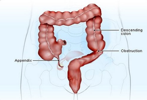
It is a tiny tubular appendage that resembles the shape of the worm. It is attached to the cecum section of the colon.

The anatomical name for appendix is vermiform. The appendix has an inner lining that produces a small amount of mucus, which flows through the open central core of the appendix.

The appendix’s wall consists of lymphatic tissue, which is a significant part of the immune system. If the appendix becomes blocked, the bad bacteria will invade and infect the appendix’s wall leading to a condition called appendicitis. (1, 2, 3)

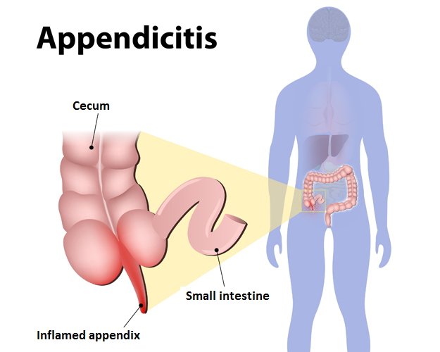
What side is your appendix on image picture photo

Photo 1: The anatomical presentation of the appendix location.
Image Source: images.onhealth.com

An image showing an inflamed appendix (appendicitis) location photo picture

Picture 2: An image showing an inflamed appendix (appendicitis).
Photo Source: www.medifee.com

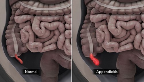
A comparison image between a normal and inflamed appendix location photo picture

Image 3: A comparison image between a normal and inflamed appendix.
Picture Source: media.licdn.com

A surgical removal of the appendix (appendectomy) location photo picture

Photo 4: A surgical removal of the appendix (appendectomy).
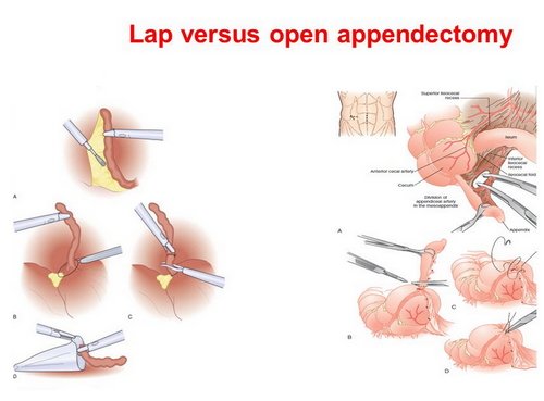
Image Source: c8.alamy.comA comparison image between minimally invasive removal of the appendix and open appendectomy location photo picture

Image 5: A comparison image between minimally invasive removal of the appendix and open appendectomy.
Photo Source: slideplayer.com

What side of the human body is the appendix on?

The appendix is located on the lower right side of the abdomen. It is the area where the small and large intestines meet. The location of the appendix is called McBurney’s point, which is about 2/3 away from the navel to the hip bone.

That is why one of the cardinal signs of appendicitis is lower right quadrant pain. Appendicitis is an extremely serious condition that warrants immediate medical intervention. If the appendix is severely inflamed, it can rupture causing intense pain on the right side of the abdomen. If not treated right away could lead to a life-threatening condition. (2, 3, 4, 5)

Clinical manifestations

  • appendix pain symptoms is usually on the right lower quadrant of the abdomen
  • nausea and vomiting
  • fever (low grade) and chills (6)
  • Initially, the appendix pain comes and goes in the middle section of the abdomen. After several hours, the pain gets severe and spreads to the lower right quadrant of the abdomen.
  • The pain gets worst if you move your limbs. Sometimes, even sneezing and coughing can cause pain.
  • Loss of appetite (6)
  • Constipation or diarrhea
  • Excess gas and a feeling of bloating
  • Abdominal swelling
  • The pain is described as dull or sharp effecting not only the abdomen but the back and rectum too. (4)

Are there other health conditions causing right lower quadrant pain?

Right lower quadrant pain is not always linked with appendicitis. There are other related conditions such as:

  • Kidney stones
  • Urinary tract infection
  • Ovulation pain
  • Inflammation of the intestines
  • Rebound tenderness (pain is felt when the lower right part of the abdomen is pushed and the pressure is released) (5, 6, 8, 9)
  • Ectopic pregnancy
  • Right-sided diverticulitis
  • Pelvic inflammatory disease

Appendix pain location

The exact location of pain is difficult to pinpoint, especially on the early onset of pain. The patient usually locate the pain in a circular motion around the center portion of the abdomen. As time passes by, the pain is localized in the right lower abdominal area, which makes it easier for the patient to locate the exact location of the pain.

The pain is dull in the navel area and becomes sharp as it moves to the lower right quadrant of the abdomen. (1, 2, 9)

Appendicitis Causes

A blockage in the appendix causes appendicitis. The blockage could be due to stool, presence of a foreign body, or cancer. The appendix can also swell during inflammatory response. (4, 6)

Appendicitis Test

There are various tests and procedures done to confirm the diagnosis of appendicitis. They are the following:

  • Physical examination – The patient’s abdominal area is checked to assess any changes that are indicator of appendicitis.
  • CT scan – A detailed image of the appendix is seen through the CT scan. You can see if the appendix is inflamed or if it has already ruptured.
  • CBC – The components of the blood is checked. An increased in white blood cells is a sign of infection.
  • Ultrasound – Sound waves are used to check for the presence of appendicitis.
  • Other diagnostic procedures – When deemed necessary, the doctor will order other special procedures such as PET scan and MRI. (5, 7, 9, 10)

Appendicitis treatment

There are various treatment modalities for appendicitis. To address any potential infection, the doctor will put the patient on antibiotics. However, antibiotics alone cannot effectively treat the underlying condition.

The typical approach for appendicitis is surgery. Appendicitis surgery is called appendectomy. It is the surgical removal of the appendix. If there is an existing tumor, there might be a need to remove a part of the colon.

The surgery is usually done through laparoscopy or keyhole surgery. It is a minimally invasive procedure. The surgeon inserts a thin tube that has a video camera and light into the abdomen. It helps the surgeon accurately view the inside structure of the abdomen. Through a monitor, the doctor can magnify the image.

A tiny instrument responds to the hand movement of the surgeon, which will facilitate the removal of the appendix. This procedure can be very costly, but has minimal blood loss because only a small incision is made. There is less scarring and the patient can fully recover in just a short period of time.

For patients who can’t afford a minimally invasive procedure, the traditional open surgery is the best option. The surgeon will create a large incision to view the inside structure of the abdomen. This is typically the surgery of choice is the appendix is already ruptured or infected, has abscess, the patient has had abdominal surgeries before, and if the patient is in her third trimester of pregnancy. (1, 3, 5, 8, 10)

Recovery period

Twelve hours post-surgery, the patient can already get up and move around. After a week, the patient is discharged from the hospital and can go back to the usual activities of daily living. If the less invasive surgery is performed such as the keyhole technique, the recovery period is pretty fast. (3, 6, 7)

Can you prevent appendicitis?

There is no prevention for appendicitis. Studies showed that people who consume a high fiber diet such as fruits and vegetables are less susceptible to appendicitis. (2, 4, 7, 8)

Appendicitis Myths

  • The appendix has no use – The truth is that the appendix helps maintain a balance of good bacteria in the body.
  • Running or jumping after meal can cause appendicitis – The truth is that appendicitis is caused by a blockage in the appendix.
  • Eating small seedy vegetables/fruits can cause appendicitis – It is highly unlikely that small seeds can cause blockage in the appendix. (4, 8, 10)

References:

  1. https://what-side-is-your-appendix-on.blogspot.com
  2. https://www.webmd.com/digestive-disorders/digestive-diseases-appendicitis
  3. https://www.medicinenet.com/appendicitis/article.htm
  4. https://www.nhs.uk/conditions/appendicitis/symptoms/
  5. https://www.medicalnewstoday.com/articles/158806.php
  6. https://www.healthline.com/health/appendicitis
  7. https://en.wikipedia.org/wiki/Appendicitis
  8. https://www.emedicinehealth.com/appendicitis/article_em.htm
  9. http://m.kidshealth.org/en/parents/appendicitis.html
  10. https://www.mayoclinic.org/diseases-conditions/appendicitis/symptoms-causes/syc-20369543
  11. https://www.onhealth.com/content/1/appendicitis_appendectomy

No responses yet

Steatorrhea

Dec 16 2017 Published by under Diseases and Conditions

Steatorrhea Definition

A human stool consists of different materials such as water, mucus, protein, fiber, salt, cell linings, bacteria, and fats.

Most of the content should be water and a little of everything mentioned above. If there is too much fat in the stool, the condition is called steatorrhea.

It is not really a medical condition but a clinical manifestation of a disease such as a malabsorption syndrome.

What causes fat in the stool? Are there any health implications? Keep on reading below. (1, 2, 3)

Steatorrhea Pictures

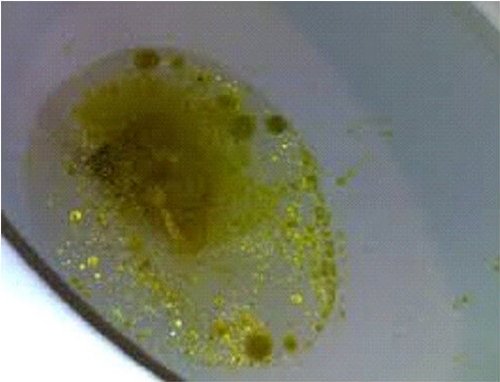
The water inside the bowl has drops of oil, which is one of the symptoms of steatorrhea image photo picture

Image 1: The water inside the bowl has drops of oil, which is one of the symptoms of steatorrhea.
Photo Source: healthfixit.com

Lactose intolerance is one of the primary causes of too much fat in the feces Steatorrhea image photo picture

Picture 2: Lactose intolerance is one of the primary causes of too much fat in the feces.
Image Source: health.facty.com

taking folic acid can actually help in the treatment, management, and prevention of steatorrhea photo image picture

Photo 3: Taking folic acid can actually help in the treatment, management, and prevention of steatorrhea.
Picture source: charlies-magazines.com

Causes of steatorrhea

  • Digestive problem – Too much fat in the stool is an indicator that the digestive system has a hard time breaking down foods.
  • Cystic fibrosis – It causes malabsorption leading to accumulation of fat in the stool.
  • Pancreatitis – It is the inflammation of the pancreas. It is the organ that secretes enzymes, which is helpful in the digestion of protein, carbohydrates, and fats in the small intestine. (3)
  • Lactose intolerance – the body lacks lactase, an enzyme used to digest the sugar in milk products.
  • Biliary atresia – the duct that carries bile from the liver to the gallbladder has a blockage.
  • Celiac disease – It is a condition in which a person has a sensitivity to gluten.
  • Whipple disease – It is a type of bacterial infection affecting the body’s ability to break down carbohydrates and fats.
  • Crohn’s disease – The gastrointestinal tract is inflamed. (4)

Steatorrhea symptoms

  • The stool looks pale and bulky.
  • The stool smells extremely foul.
  • The stool tends to float because of high gas content.
  • If you look at the water inside the bowl, you will notice drops of oil.
  • The patient complains of indigestion and abdominal cramps.
  • Loose stool and weight loss (3, 4, 5)

Steatorrhea diagnosis

If your stool is greasy and extremely foul, then you need to inform your doctor. The doctor will thoroughly assess your condition and will order various tests to accurately diagnose your condition. The number of fat globule in the stool sample will be measured.

The normal result should be less than 50 neutral fat globules and lower than a hundred fatty acid fat globules. Another way to test steatorrhea is collecting stool samples for at least four days and the amount of fat in the stool is checked every single day. A normal fat content should be around 2 to 7 grams/24 hours for adults.

That fat content should not be more than 20% of the solid stool sample. For infant’s stool, it should be less than 1 gram/24 hours. Breastfed babies’ stool must contain at least 10% to 40% of fat in the stool. On the other hand, bottle-fed babies usually have 30% to 50% fat in the stool. (2, 5, 6)

An abdominal x-ray, CT scan, and ultrasound can be ordered to check for pancreatic and small intestine diseases, which are the leading cause of too much fat in the stool. If the doctor suspects a small intestinal disease, the following diagnostic procedures are ordered:

  • D-xylose absorption – It will test the function of the proximal small bowel. D-xylose is a type of sugar.
  • Schilling tests (I and II) and bile acid breath tests – These tests will detect bacterial overgrowth.
  • Barium study – It will help detect any abnormalities in the small bowel. (7)

Steatorrhea treatment

Treatment for steatorrhea is focused on treating the underlying cause. To come up with the most effective treatment, a reliable and accurate diagnosis should be made. The treatment modalities usually include the following:

  • Diet modification – If too much fat in the stool is diet-related, the best remedy is to modify the diet. If you have lactose intolerance, you should avoid milk and dairy products. If you have a celiac disease, you should avoid foods containing wheat and gluten.
  • Medications – If it is caused by an underlying medical condition, then it should be treated using the right medication. The primary cause of too much fat in the stool should be addressed first. For an instance, a patient suffering from chronic pancreatitis should take potent enzymes (pancreatic enzyme replacement). It helps reduce the passage of bulky stools. (5, 8, 9)

What can you do to prevent steatorrhea?

Some of the causes of steatorrhea are due to human lifestyles such as too much fat in the diet and nutritional deficiencies. Hence, it is important to take good care of the body. If you are the type of person who loves to eat fatty foods, then you should cut back the portion of what you eat.


Always make it a habit to check the nutritional label when shopping for foods. If you are deficient in certain nutrients, then you should take nutritional supplements. They are available at the leading stores.

Steatorrhea can be short-term or long-term. If steatorrhea is short-term, then it is most likely due to diet or GI infection. However, if you continue to notice too much fat in your stool, it could be an indicator of a serious underlying condition. Therefore, it is important to consult your doctor the soonest time possible. (2, 5, 7, 8)

References:

  1. https://en.wikipedia.org/wiki/Steatorrhea
  2. https://www.healthline.com/health/steatorrhea
  3. https://www.diagnose-me.com/symptoms-of/steatorrhea.php
  4. https://www.healthgrades.com/symptoms/steatorrhea
  5. http://healthfixit.com/steatorrhea/
  6. http://www.primehealthchannel.com/steatorrhea-pictures-definition-symptoms-causes-and-treatment.html
  7. http://pathwaymedicine.org/steatorrhea
  8. https://www.belmarrahealth.com/floating-poop-steatorrhea-understand-why-your-stool-floats/
  9. https://medicalfoxx.com/steatorrhea.html
  10. https://www.medigoo.com/articles/steatorrhea/

No responses yet

Mucus In Urine

Dec 16 2017 Published by under Diseases and Conditions

A mucus is a thick and slimy substance commonly found in the nose, throat, mouth, and urinary tract. It coats and moistens these areas of the body.

Being in the urinary tract, it is just normal to have a small number of mucus in the urine. However, too many mucus in the urine isn’t normal. (1, 2)

A urine sample showing epithelial cells Mucus in urine image photo picture

Image 1: A urine sample showing epithelial cells.
Photo Source: www.newhealthadvisor.com

A urine sample of a child with a large number of mucus Mucus in urine image photo picture

Picture 2: A urine sample of a child with a large number of mucus.
Image Source: upladdy.com


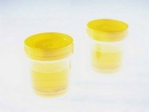
A urine sample of a patient that appears cloudy, which is indicative of excess amount of mucus image picture photo

Photo 3: A urine sample of a patient that appears cloudy, which is indicative of excess amount of mucus.
Picture Source: cdn.grapegate.com

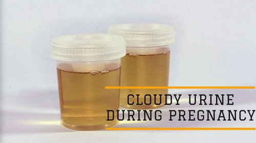
A urine sample of a pregnant woman Mucus in urine image photo picture

Image 4: A urine sample of a pregnant woman.
Photo Source: www.mypostpartumwellness.com

Symptoms

  • High number of mucus in the urine during urinalysis
  • Abdominal pain (3)
  • Pain and discomfort when urinating
  • Fever
  • Pain during sexual intercourse
  • Foul-smelling urine (2)

Reasons for mucus in urine

  • Urinary tract infection – It is caused by bacteria. The bacteria attack the bladder and other parts of the urinary system. One of the primary signs of UTI is discomfort in urinating.
  • Irritable bowel syndrome – There is an excessive production of mucus in the large intestine. The excess mucus escapes in the urine making the urine cloudy or thick in appearance. It is what causes the urine to be thick.
  • Sexually transmitted disease – The organisms that cause sexually transmitted disease stay in the urethra. Once the urine passes through it the mucus gets flushed out.
  • Ulcerative colitis – It is a type of inflammatory bowel disease wherein there is an ulceration in the mucus membrane leading to an abnormal increase in the production of mucus. The excess mucus leaks in the urinary system.
  • Obstruction in the urinary tract – Mucus in the urine can sometimes cause by urinary tract obstruction. Examples are patients with kidney stones. (3, 4, 5, 6)

Mucus urine test

A urinalysis or urine test is ordered to check the components of urine. It can detect whether there is too much mucus in the urine. A mucus urine test is a special procedure that is ordered if you are demonstrating the symptoms of urinary tract infection such as painful urination, frequent urination, foul smelling urine, cloudy urine, fatigue, and weakness.

During the mucus urine test, a urine sample is collected using the clean catch method. It is just normal to see small to moderate mucus threads in the urine. If there is a large amount of mucus threads in the urine, then it could mean that you are suffering from a serious health condition such as UTI, sexually transmitted disease, kidney stones, irritable bowel syndrome, and bladder cancer. (6, 7, 8)

What does squamous epithelial cells in urine mean?

Epithelial cells line the surface of the body such as the skin, urinary tract, blood vessels, and vital organs. Having a small amount of epithelial cells in the urine is normal. However, if the number grows out of control it could indicate an underlying medical condition such as kidney disease.

It could also indicate that the urine sample is contaminated. Normally, there is between 15 and 20 squamous epithelial cells/hpf. Anything more than that is abnormal. (8, 9)

Mucus in urine during pregnancy

A mucus in urine during pregnancy is common because a pregnant women is at risk for urinary tract infection. This is primarily due to the changes in the urinary tract. Anatomically speaking, the uterus sits on top of the bladder.

As it grows during pregnancy, it blocks the draining of the urine leading to an infection and overgrowth of mucus threads. (2, 5, 10)

Mucus in Urine Treatment

If there is an excess number of mucus in the urine, medical attention should be given right away. Some of the treatment modalities used by health care professionals include the following:

  • Intravenous therapy – The patient is put on intravenous fluids to help flush out the mucus from the urine.
  • Increase fluid intake – The patient is highly encourage to drink at least 12 glasses of water a day to promote urination thereby facilitating the flushing of mucus from the urine.
  • Antibiotic therapy – the doctor will start you on antibiotic therapy to treat an underlying infection.
  • Proper hygiene – It is important to observe proper hygiene so that you can avoid catching urinary tract infection. It also helps prevent the formation of mucus in the urine.

A mucus in urine in small to moderate amount isn’t really a cause of concern. However, too mucus in the urine is an indicator that something is wrong in your body. Hence, it is important to consult your doctor right away if you are experiencing any of the symptoms mentioned above.

Not treating the underlying condition in a timely manner could lead to irreversible damage. (1, 4, 6, 7, 9)

References:

  1. https://medlineplus.gov/labtests/mucusinurine.html
  2. https://www.healthline.com/health/mucus-in-urine
  3. https://www.belmarrahealth.com/mucus-urine-common-causes-treatment/
  4. https://www.doctorshealthpress.com/health-articles/mucus-in-urine-causes-natural-remedies/
  5. http://www.newhealthadvisor.com/Mucus-Threads-in-Urine.html
  6. http://symptomscausestreatment.com/mucus-in-urine-symptoms-causes-treatment.html
  7. http://www.actforlibraries.org/mucus-threads/
  8. https://healthsaline.com/mucus-in-urine.html
  9. https://medinfo.org/mucus-in-urine
  10. https://www.ihealthblogger.com/mucus-in-urine/

No responses yet

Thyromegaly

Dec 10 2017 Published by under Diseases and Conditions

The thyroid gland is one of the most important parts of the body. It is the gland that secretes thyroid hormones, an essential hormone used for various bodily processes.

A thyroid gland has a shape similar to a butterfly. It is found below the Adam’s apple. A healthy thyroid gland regulates the right amount of thyroid hormone.

If the thyroid gland grows bigger than it is supposed to, it could result in a thyromegaly or commonly called goiter. (1, 2)

Thyromegaly ICD 9 : 240.9

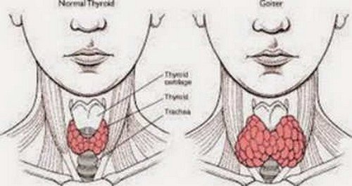
A comparison image between a healthy-normal thyroid and a swollen thyroid thyromegaly image photo pictures

Image 1: A comparison image between a healthy/normal thyroid and a swollen thyroid (thyromegaly).
Photo Source: healthosphere.com

A doctor performing physical exam in a patient suspected of an enlarged thyroid thyromegaly image picture photo

Picture 2: A doctor performing physical exam in a patient suspected of an enlarged thyroid.
Image Source: 3.bp.blogspot.com

An image showing a normal thyroid, a thyroid that has undergone a partial thyroidectomy, and a subtotal thyroidectomy thyromegaly photo image picture

Photo 3: An image showing a normal thyroid, a thyroid that has undergone a partial thyroidectomy, and a subtotal thyroidectomy.
Picture Source: khalidalomari.weebly.com

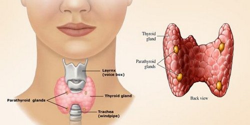
An image showing the anatomy of the thyroid gland thyromegaly picture images photo

Picture 4: An image showing the anatomy of the thyroid gland.
Photo Source: www.organsofthebody.com

Thyromegaly Symptoms

  • Swelling of the neck
  • Breathing difficulty
  • Discomfort in the neck area when eating, drinking, or swallowing
  • Stiffness in the throat area
  • The voice becomes hoarse and accompanied by cough
  • The neck is swollen
  • Headache and dizziness (1, 2, 3)

Thyromegaly Causes

  • Heredity – Thyromegaly runs in the family. If any of your family members have a thyromegaly, there is a tendency that it could happen to you too.
  • Diet – If your diet lacks iodine, then you are prone to having thyromegaly.
  • Side effects of medications – Some drugs such as cobalt and lithium can have drastic effect to the thyroid.
  • Old age – The risk of thyroid problem increases as the person grows older.
  • Medical conditions – Some medical conditions increase the chance of having thyromegaly such as Grave’s disease. It causes hyperthyroidism wherein the immune system of the body attacks the thyroid gland. Other health conditions that could lead to thyromegaly are thyroiditis (inflammation of thyroid gland) and thyroid cancer. (3, 4, 5, 6)

Varieties of thyromegaly

  1. Hyperthyroid – The hormones produced by the thyroid gland are more than the usual causing the thyroid gland to enlarge.
  2. Euthyroid – It does not affect the thyroid gland’s normal functions. The mass grows but is not cancerous.
  3. Hypothyroid – The thyroid gland is not producing the right amount of thyroid hormone. It is categorized into three:
  1. Diffuse goiter- the thyroid gland is entirely covered by big masses.
  2. Non-toxic – It reduces the functions of the thyroid gland.
  3. Toxic goiter – The mass is bigger than the usual. (5, 6,, 7)

Who are at risk?

  • For unknown reasons, women are more prone to thyromegaly than men.
  • Older people (more than 40 years old) are prone to thyromegaly, although young people can also have thyromegaly.
  • People who take medications containing high doses of cobalt and lithium.
  • People with family history of goiter.
  • People who are exposed to radioactive materials for a long period of time.
  • Some infections can cause goiter.
  • Pregnancy can sometimes affect the functions of the thyroid gland. (7, 8, 9)

Diagnosis

  • Physical examination – The doctor will thoroughly check the throat area for swelling and inflammation. A doctor who has a vast experience in handling thyromegaly cases can easily detect one by just merely doing a physical examination.
  • Thyromegaly ultrasound – It is a non-invasive procedure that gives a clear view of the thyroid gland.
  • TSH Test – It checks the level of thyroid stimulating hormone in the blood.
  • T3 Test – It check the level of triiodothyronine in the blood.
  • T4 test – It checks the level of thyroxine in the blood. (2, 5, 10)

Thyromegaly Treatment

If the thyromegaly is mild and caused by low iodine level in the body, the doctor will put the patient in an iodine supplement. If the thyromegaly is severe causing other discomforting symptoms, then the doctor will put the patient in a course of treatment which includes suppression therapy.

This is to reduce the secretion of levothyroxine. Ethanol infusion can also help in the treatment of non-cancerous lump in the thyroid. (1, 3, 4)

Diet plays a very important role in improving the function of the thyroid gland. There are foods you should eat and foods you should completely avoid. They include the following:

Foods to avoid

  • Cruciferous vegetables like cabbage, broccoli, cauliflower, kale, and Brussels sprout, to name a few.
  • Soy products like soy beans, soy milk, tofu, and anything that has a soy in its label.
  • Peanuts, spinach, peaches, and strawberries. (4, 5)

Foods to eat

  • Marine fish or fish that comes from the ocean.
  • Shellfish such as shrimps, oysters, mussels, and clams.
  • Milk and yogurt
  • Eggs
  • Use iodine treated salt (iodized salt) (9, 10)

Are surgical interventions necessary?

A surgery is needed if the patient develops malignancy or cancer in the thyroid gland. Surgery is the last resort. It should only be done if the radioactive iodine therapy didn’t work. The doctor will perform a surgery because of the following reasons:

  • A nodule is present, which is an indicative of a thyroid cancer.
  • Your diagnosis is confirmatory of a thyroid cancer.
  • The nodule in the thyroid is causing discomfort such as difficulty swallowing and compression of the trachea. The mass in the throat can be unsightly, which makes people with thyromegaly want to remove it. (3, 6, 7)

Thyroidectomy is the surgical removal of the thyroid gland. It is categorized into two: partial and total thyroidectomy. A is a procedure in which the nodule is excised directly is called open thyroid biopsy. If only one love of the thyroid is removed, the procedure is called hemi-thyroidectomy.

There are instances when only a bridle of the thyroid tissue between the two loves is removed. This procedure is called isthmusectomy. If most or all of the thyroid tissue is removed, the procedure is called total or near total thyroidectomy.

A lot of people are not aware that they are suffering from thyromegaly. Hence, it is important to be very cautious on the signs and symptoms of goiter. Have yourself regularly checked to find out if you have been suffering from an underlying condition and that it should be treated at an early stage.

Thyromegaly is a serious condition which could lead to serious and fatal complications if left untreated. (5, 9, 10)

References:

  1. http://diseasespictures.com/thyromegaly/
  2. https://www.doctorshealthpress.com/general-health-articles/thyromegaly-goiter-causes-treatment/
  3. https://healthosphere.com/thyromegaly/
  4. https://medinfo.org/thyromegaly-101-what-you-should-know.html
  5. https://medicalpoint.org/thyromegaly/
  6. http://healthinstant.net/thyromegaly-symptoms-causes-treatment/
  7. http://diseasedefinition.info/thyromegaly-definition-causes-signs-symptoms-treatment-and-prevention/
  8. https://safesymptoms.com/what-is-thyromegaly/
  9. http://healthncare.info/thyromegaly-enlarged-thyroid-symptoms-causes-treatment/
  10. https://allhealthpost.com/thyromegaly/

No responses yet

© 2023 Healthooze.com. All Rights Reserved. Privacy Policy