Archive for July, 2018

Bruised Ribs

Jul 22 2018 Published by under Uncategorized

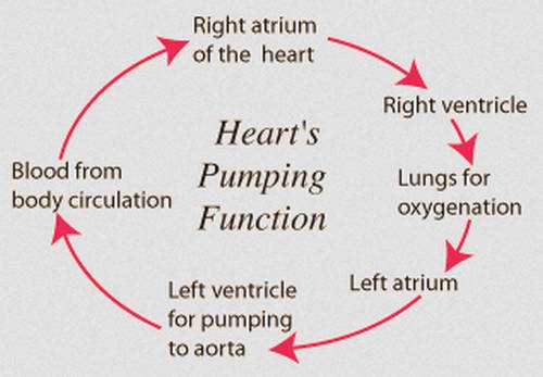
The ribs are thin bones that enclose the vital organs of the body such as the heart, lungs, and chest cavity. Aside from protecting the important organs, ribs also aid in breathing.

However, there are instances when the structure and integrity of the ribs are compromised. A trauma to the chest can lead to a bruised rib. (1, 2)

A severe bruising of the ribs image photo picture

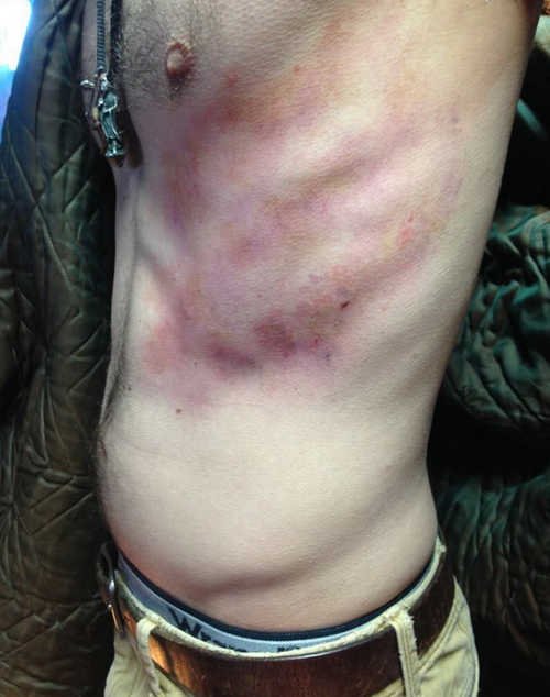
Image 1: A severe bruising of the ribs.
Picture Source: i.pinimg.com

A bruised rib as seen on the patient’s skin discoloration, tenderness, and inflammation image photo picture

Picture 2: A bruised rib as seen on the patient’s skin discoloration, tenderness, and inflammation.
Photo Source: www.epainassist.com

Who are at risk for a bruised rib?

People who are fond of playing contact sports are prone to have bruised ribs such as those who play football, hockey, rugby, wrestlers, boxers, and lacrosse players. A vehicular accident can also put you at risk for a bruised rib.

Other possibilities include falling from a high place or a heavy object fell on your chest. Performing strenuous activities, lifting heavy objects, and excessive coughing can all lead to bruised ribs. (1, 2, 3)

Signs and symptoms

  • Pain in the chest that gets worse with inhalation. The pain gets intense if you try to sneeze, laugh, or a cough.
  • Simple activities like bending or moving can trigger chest pain.
  • Muscle spasm in the rib cage area.
  • Tenderness and swelling around the bruised rib.
  • There is a visible bruising on the skin. (2, 3, 4)

Diagnosis

An imaging study of the chest wherein there is an obvious rib bruising secondary to excessive coughing image photo picture

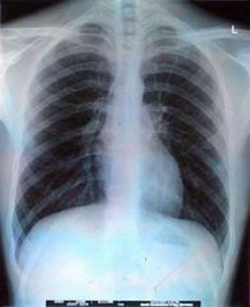
Photo 3: An imaging study of the chest wherein there is an obvious rib bruising secondary to excessive coughing.
Image Source: userfiles.steadyhealth.com

An imaging study of a bruised rib image photo picture

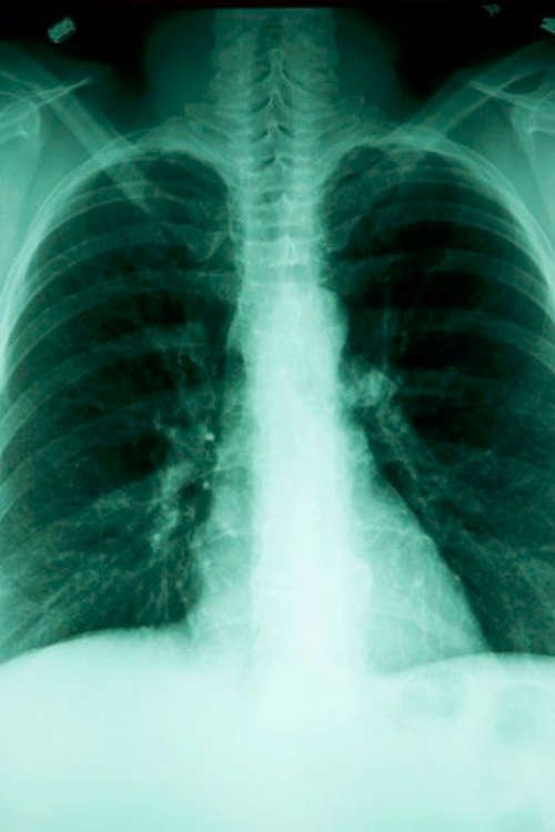
Image 4: An imaging study of a bruised rib.
Picture Source: img.aws.livestrongcdn.com

Physical examination

The doctor will perform a thorough physical examination to accurately assess the symptoms of the patient. Your breathing will be carefully assessed to note if the functions of the lungs are affected.

Any bruising on the skin on the chest and back will be assessed too.

X-ray

It is done to check the condition of your chest and to detect any issues that cannot be detected by simple physical examination. The doctor will order for a lateral and anteroposterior x-ray to check for the structure of the rib cage and to rule out rib fracture.

CT scan

This is a more thorough and accurate imaging study which will help differentiate a bruised rib from a break/fracture. It is performed to determine the number of ribs involved. It also helps detect the extent of the injury.

MRI

A bruised rib can be easily detected under the magnetic resonance imaging study.

Bone scan

It is a more advanced procedure that will not only detect a bruised rib but a broken rib as well.

Angiography

It is performed to assess for blood vessel injury as the first and second ribs are closed to the important blood vessels.

Arterial blood gas

It can detect the extent of bruising, especially the lungs, which could affect the lung’s ability to transport oxygen.

Complete blood count

It helps determine blood loss and the general condition of the essential blood components. (4, 5, 6, 7)

Bruised ribs Treatment

The treatment for a bruised rib is focused on pain management. The patient should have adequate rest and to avoid activities that can further aggravate the pain. To manage the pain, the patient is put on pain reliever and anti-inflammatories such as acetaminophen, aspirin, ibuprofen, and the likes.

Aside from taking a pain reliever, you can also manage the pain through cold compress. It is best to apply cold the earliest time possible for about 20 minutes for at least three times a day. It will help alleviate the pain, reduce inflammation, and discomfort.

A bruised rib can significantly affect your breathing. It forces you to take more shallow breaths. This is one of the reasons why your doctor will put you on a pain medication. In some instances, a long-lasting anesthetic injection is introduced near the bruise site to alter the body’s pain signals.

The doctor strongly advised the patient to undergo a respiratory therapy so as to learn breathing techniques that can significantly help reduce the pain when breathing and at the same time supplying the much-needed amount of air in the lungs. (6, 7, 8, and 9)

How long will it take to heal bruised ribs?

The time it takes for the bruised ribs to heal depends on the extent and severity of bruising. Usually, it takes a few weeks to months. However, the recovery period is long if several ribs are affected. Usually, the pain subsides after a few weeks, which means that your bruising is healing.

If the pain is still there and gets worse as time passes by, then you need to tell your doctor. It needs to be re-evaluated to make sure that it is just a bruise and not an underlying rib fracture. (5, 7, 9)

When should you call out for help?

You need to thoroughly assess your condition and make sure you immediately call out for help if anything gets worse such as:

  • Difficulty breathing/shortness of breath.
  • Rib pain, especially when breathing or coughing.
  • Bruising or swelling around the rib area.
  • The pain gets worse as time passes by. (1, 2, 3)

What to keep in mind?

A bruised rib is something that should not be taken lightly. It may sound like a simple problem but if it affects several ribs, the integrity and function of your lungs will be greatly affected. You need to strictly follow your doctor’s advice to hasten the recovery period.

Take the pain medication as prescribed. You have to understand that the first few weeks of healing can be extremely painful for the patient. If the pain does not subside using pain medication, then you need to inform your doctor right away.

A higher dose of pain medication might be needed such as a nerve block to numb the area between the ribs. The best way to safeguard yourself from bruised ribs is to observe preventive measures such as wearing protective gear when engaging in contact sports. (2, 7, 9, 10)

References:

  1. https://www.healthline.com/health/bruised-ribs
  2. https://www.nhs.uk/conditions/broken-or-bruised-ribs/
  3. https://www.your.md/condition/rib-injuries/
  4. http://www.sportsinjuryclinic.net/sport-injuries/chest-abdomen-pain/bruised-ribs
  5. http://www.coreperformance.com/knowledge/injury-pain/bruised-ribs-what-you-need-to-know.html
  6. https://www.belmarrahealth.com/bruised-ribs-symptoms-causes-natural-remedies/
  7. https://allhealthpost.com/bruised-ribs-symptoms-causes/
  8. http://www.md-health.com/Bruised-Ribs.html
  9. https://www.physioadvisor.com.au/injuries/upper-back-chest/rib-contusion/
  10. https://www.physiocheck.co.uk/condition/80/bruised-or-broken-rib

No responses yet

Anatomical Snuff Box

Jul 21 2018 Published by under Uncategorized

What is Anatomical snuff box?

It is a triangle-shaped depression on the lateral part of the dorsum of the hand. What is the medical term for snuff box? In the medical field, it is called radial fossa. In the medical term, a fossa pertains to a hollow depression/semi-flattened surface.

The snuff box is located at the level of the carpal bones. If you try to locate the snuff box, all you have to do is to extend your thumb. It is fondly called the snuff box because it was used to hold snuff (ground tobacco) as it inhaled through the nose. (1, 2, 3)

The borders of the anatomical snuff box image photo picture

Image 1: The borders of the anatomical snuff box.
Picture Source: cdn1.teachmeseries.com

The image showing how to properly and accurately locate the anatomical snuff box image photo picture

Picture 2: The image showing how to properly and accurately locate the anatomical snuff box.
Photo Source: medicine.academic.ru

Anatomical snuff box borders

  1. Ulnar – It is the medial border specifically the extensor pollicis longus tendon.
  2. Radial – it is the lateral border, which includes the abductor pollicis longus and extensor pollicis brevis tendons.
  3. Proximal – It includes the radius’ styloid process. (2, 3, 4)

The anatomical snuff box along with other important structures image photo picture

Photo 3: The anatomical snuff box along with other important structures.
Image Source: pbs.twimg.com

What are the contents of the anatomical snuff box?

The primary contents of the anatomical snuff box are:

Radial artery

It crosses the anatomical snuff box’s floor in an oblique way leading to the extensor tendons. If you place your two fingers near the anatomical snuff box, you can easily palpate the radial pulse.

Branches of radial nerve

The terminal branches of the radial nerve run across the upper parts of the anatomical snuff box.

Cephalic vein

It crosses the anatomical snuff box. (4, 5, 6)

The anatomical snuff box location

The anatomical snuff box is strategically located on the thumb side of the hand or in the medical term the lateral/radial part of the hand. To locate your snuff box, you have to do the following:

  • Spread all the five fingers out/make your hands look wide.
  • Notice a small indentation on the wrist; that is your anatomical snuff box.
  • Try to give someone a thumbs up and the snuff box will be visible at the wrist joint. (2, 4, 5, 6)

Anatomical snuff box from a skeletal standpoint

From a skeletal standpoint, the snuff box is located over the carpals; the group of bones found in the wrist. There are eight uniquely shaped bones in the wrist.

The carpal bone, specifically the scaphoid bone serves as the floor of the anatomical snuff box. It is the foundation of the anatomical snuff box. (6, 7, 8)

The anatomical snuff box from a muscular standpoint

The anatomical snuff box has two thin and chord-like structures forming the snuff box’s triangular shape creating the divot.

The medial border has the tendon of the extensor pollicis longus muscle. The lateral border of the snuff box has the tendons of the muscles pollicis longus (abductor) and pollicis brevis (extensor). (7, 8)

Nerves and blood supply

The anatomical snuff box has nerves and arteries supplying the area with blood and making it responsible for various sensation.

  • Radial artery – It supplies blood to the essential parts of the wrist and hand.
  • Radial nerve – It provides sensation and functions. (6, 7, 8)

Anatomical snuff box clinical significance

Scaphoid fracture

The scaphoid together with the radius articulate forming the wrist joint. If a person sustains a fall on an outstretched hand, it could cause a severe blow to the wrist leading to excruciating pain. A localized pain in the anatomical snuff box is most likely due to scaphoid fracture.

It is something that should not be taken lightly because any forms of fracture to the scaphoid could greatly affect the blood supply to the proximal part of the anatomical snuff box. Failure to address the problem right away could lead to avascular necrosis and arthritis.

Idiopathic radial artery aneurysm of the anatomical snuff box

It is an extremely rare condition and is linked to trauma that is penetrating in nature. (8, 9, 10)

The anatomical snuff box-scaphoid fracture as manifested by wrist pain image photo picture

Image 4: The anatomical snuff box/scaphoid fracture as manifested by wrist pain.
Picture Source: acuclinic.com.au

Swelling and tenderness are the cardinal signs of anatomical snuff box fracture image photo picture

Picture 5: Swelling and tenderness are the cardinal signs of anatomical snuff box fracture.
Photo Source: encrypted-tbn0.gstatic.com

Anatomical snuff box injury

If you are suspecting an anatomical snuff box injury, a thorough physical examination and diagnostic procedure should be performed.

Physical examination

  • Compare the injured wrist with an uninjured wrist.
  • Check for signs of tenderness on examination as it is one of the cardinal signs of scaphoid fracture.
  • Check for the tenderness of the scaphoid tubercle. The doctor extends the wrist of the patient with one hand and applies pressure to the tuberosity using the opposite hand. If no tenderness is felt, then the patient does not have a scaphoid fracture.
  • Pain with an anatomical snuff box compression test. The patient’s thumb is compressed along the line of the first metacarpal.
  • Pain in the snuff box as the wrist is pronated secondary to the ulnar deviation. (1, 6, 9, 10)

Imaging studies

Plain radiographs

To evaluate a fracture in the anatomical snuff box, a radiographic view; anteroposterior, lateral, and oblique should be done.

MRI (magnetic resonance imaging)

It is ordered to detect anatomical snuff box fracture the earliest possible time because MRI is specific and sensitive.

Ultrasonography

A high spatial resolution ultrasonography is performed to identify occult scaphoid fracture.

Bone scintigraphy

It is done to detect occult fracture of the anatomical snuff box and other bones. It is more accurate and cost-effective compared to other imaging studies. (2, 4, 6, 7)

Anatomical snuff box fracture Treatment

The type of treatment for anatomical snuff box fracture depends on the extent and severity of the fracture. For someone with a suspected anatomical snuff box fracture and negative radiograph, the best approach is to apply a short arm thumb spica and the fracture needs to be evaluated in 10 to 14 days.

It is important to apply a cast the right way as failure to do so can worsen the patient’s condition. A bone scintigraphy might be needed should the patient opted for an alternative treatment method. (2, 5, 9)

References:

  1. https://en.wikipedia.org/wiki/Anatomical_snuffbox
  2. http://teachmeanatomy.info/upper-limb/areas/anatomical-snuffbox/
  3. https://radiopaedia.org/articles/anatomical-snuff-box
  4. https://www.physio-pedia.com/Anatomical_snuff_box
  5. https://study.com/academy/lesson/what-is-the-anatomical-snuffbox-muscles-anatomy.html
  6. https://www.ncbi.nlm.nih.gov/books/NBK482228/
  7. https://www.medicinenet.com/script/main/art.asp?articlekey=13291
  8. http://sketchymedicine.com/2011/12/the-anatomical-snuff-box-and-dequevains-tenosynovitis/
  9. https://www.howtorelief.com/anatomical-snuffbox/
  10. http://masterofmedicine.com/anatomical-snuffboxboundaries-and-contents/

No responses yet

Best baby food maker

Jul 19 2018 Published by under Uncategorized

A lot of people are heading towards healthy living. It makes them choose better and healthier foods. In fact, parents are now meticulously choosing the foods they give to their babies. They no longer buy readymade baby foods. Many of them would rather prefer fresh and healthy baby foods.

Some people think that it is difficult and time-consuming for preparing baby foods from scratch but the truth is that it is easy. You just need to make sure you have the right tools and cooking equipment. When it comes to creating baby food from scratch, you need to have the best baby food maker.

There are many types of baby food maker available on the market today. Some of them are used purely for preparing baby foods while others can be used for various purposes. The price varies depending on the features and quality of the baby food maker.

The baby food maker costs more than buying readymade baby food but you will realize that you will eventually save money in the long run. Readymade baby foods are a bit expensive, especially those in the jars.

Baby food manufacturers claim that their product is 100% healthy and preservative-free. However, there is really no guarantee unless you prepare your baby foods in your own kitchen. (1, 2, 3, 4)

Baby food maker Types

All around food maker

it blends and steam baby foods. The design varies depending on the brand and model. Some have one bowl wherein you can steam and blend the baby food. Others have two bowls, one for steaming and one for blending.

Blender only

it has one function and that is to blend foods. If you are only preparing fruits, then you can have it blend directly. However, if you are going to give vegetables to your baby, then you need to pre-cook it first before blending. (4, 5, 6)

Let us take a look at the best baby food maker 2018. They are the following:

Beaba Babycook

The standard baby food maker from the Beaba Babycook company image photo picture

Image 1: The standard baby food maker from the Beaba Babycook company.
Picture Source: www.beaba.com

This baby food maker is one of the must-have tools, especially for new parents. It can be used in preparing different kinds of foods for your baby such as vegetables, fruits, fish, and meat. It can steam, blend, defrost, and reheat foods.

What’s good about this baby food maker is its one-hand operation. So, even if you are carrying your baby, you can prepare your baby’s foods in just 15 minutes.

Now, that’s a breeze! If you are doing a lot of things all at once such as attending to your baby while preparing food, you don’t have to worry as the baby food maker comes with a bell and a whistle. The design is sleek and compact and will definitely not eat much of space. (4, 5, 6, 7)

Magic Bullet Baby Bullet Baby Care System

A truly magical way to prepare fresh baby foods image photo picture

Picture 2: A truly magical way to prepare fresh baby foods.
Photo Source: i5.walmartimages.com

Preparing baby food has never been this easy and convenient. The magic bullet baby bullet baby care system comes with a 20-piece kit of accessories which will definitely take baby food cooking to a whole new level. It is a steamer and blender in one.

It works the same way as the food processor. It has trays and storage cups with dials. You can easily take note of the production date. As you know, parents want to make sure that the foods they are feeding their babies are fresh. (6, 7)

Cuisinart BFM-1000 Baby Food Maker and Bottle Warmer

A sturdy baby food maker with only one container for steaming and pureeing image photo picture

Photo 3: A sturdy baby food maker with only one container for steaming and pureeing.
Image Source: images-na.ssl-images-amazon.com

This baby food maker steams and purees food in the same container. It comes in handy and busy parents can prepare baby foods in an instant. It enables you to prepare everything in a single container which lets you save time and energy.

It also comes with a bottle warmer which makes re-heating of food a breeze. Cuisinart baby food maker comes with a huge bowl that can accommodate up to four cups. So, if you are preparing big batches of baby food, then this food maker is a perfect choice. (6, 7, 8)

Sage Spoonfuls Baby Food Maker

A sleek baby food maker perfect for moms on a tight budget image photo picture

Image 4: A sleek baby food maker perfect for moms on a tight budget.
Picture Source: hangrybabies.com

It is one of the most affordable baby food makers in the market. Although it is cheaper than the rest, it does not disappoint you when it comes to functions and features. It comes with a food processor and an immersion blender.

You have the option to buy its glass container, which is a perfect alternative to plastic baby food storage. It is microwave and dishwasher safe. (8, 9)

Babymoov Nutribaby

An all-around baby food maker image photo picture

Picture 5: An all-around baby food maker.
Photo Source: image.darty.com

It is a single tool with a multitude of uses. It is a highly recommended baby food maker for all parents. It steams, blends, heats, sterilizes, warms, and defrosts foods. One of the selling points of Babymoov

Nutribaby is its ability to steam two separate foods with varying cooking times. The steaming bowl is extra-large which is perfect for preparing a large batch of foods. Busy moms can surely find this tool helpful as it will enable them to prepare fresh baby food for the whole week.

The blender can accommodate up to six cups of fresh foods. Warming the food is not a problem as it can warm up three bottles all at once.

With baby food maker, you can now say goodbye to daunting and tiring baby food preparation. In just a few minutes, you can have your own baby food prepared in your own kitchen in just a few minutes. No need to run to the grocery store every time you run out of baby foods. All you need are fresh fruits and vegetables and a handy tool, which is the baby food maker.

There are a lot of baby food maker brands and styles to choose from. The ones mentioned above are just some of the best options. There are a lot more. When making your choice, you have to take into account your baby’s needs, your schedule, and most importantly, your budget. (3, 8, 9, 10)

References:

  1. https://www.babylist.com/hello-baby/best-baby-food-makers
  2. https://parenting.reviewed.com/best-right-now/the-best-baby-food-makers-and-mashers
  3. https://mommyhood101.com/best-baby-food-makers-2018
  4. https://www.thebump.com/a/best-baby-food-maker
  5. https://www.verywellfamily.com/best-baby-food-makers-4151951
  6. https://www.rookiemoms.com/best-baby-food-maker/
  7. https://www.momtricks.com/baby-food-makers/
  8. https://parent.guide/best-baby-food-maker/
  9. https://www.motherandbaby.co.uk/shopping-and-reviews/shopping-ideas/the-best-baby-food-makers-and-blenders
  10. https://www.whattoexpect.com/baby-products/nursing-feeding/best-baby-food-makers

No responses yet

Human Heart Diagram

Jul 19 2018 Published by under Uncategorized

The heart is one of the vital organs in the human body. It pumps blood throughout the body and supplies nutrients and oxygen to the tissues. Oxygenated blood is needed by the body to be active and perform its vital functions. The heart beats about 72 times per minute.

The heart is a muscle that expands and contracts. The pumping of the heart is referred to as the cardiac cycle and each cycle lasts eight-tenths of a second. (1, 2, 3)

The clinical presentation of the human heart image photo picture

Image 1: The clinical presentation of the human heart.
Picture Source: img.purch.com

The different parts of the human heart image photo picture

Picture 2: The different parts of the human heart.
Photo Source: www.worldinvisible.com

The human heart highlighting the four heart valves image photo picture

Photo 3: The human heart highlighting the four heart valves.
Image Source: www.cardiosmart.org

The Anatomy of the heart

  • Measurement – The size of the heart is the same as the size of the large fist. It weighs between 10 to 12 ounces in men and 8 to 10 ounces in women.

Chambers – There are four chambers in the heart; two atria (upper chamber) and two ventricles (lower chambers).

  • Right atrium and right ventricle – consist the right side of the heart.
  • Left atrium and left ventricle – consist the left side of the heart.
  • Septum – it is a wall muscle that separates one side of the heart to the other.
  • Pericardium – it encases, protect, and anchor the heart inside the chest.
  • Pericardial fluid – it lubricates the heart during contractions.
  • Outer wall – The outer wall of the heart has three layers:
  • Epicardium – the outermost wall layer.
  • Myocardium – the middle layer containing the muscle used for contraction.
  • Endocardium – inner layer of the heart; the one that gets in contact with the blood. (2, 3, 4, 5)

Heart valves – The heart has four valves:

  • Mitral valve and tricuspid valve (Atrioventricular valves) – They are found between the atria and the ventricles. They control the flow of blood from the atria to the ventricles.
  • Aortic valve and pulmonary valve (semilunar valves) – They are found between the ventricles and the major blood vessels. They control the flow of blood out of the ventricles.

Conduction of the heart 

The heart has the ability to set its own rhythm and signal needed to coordinate the rhythm.

  • Sinoatrial node – It produces an electric pulse causing the heart to contract. It is the pacemaker of the heart. It is found in the wall of the right atrium just below to the superior vena cava.
  • Atrioventricular node – It is located in the right atrium, just below the interatrial septum. It picks up the signal sent by the sinoatrial node and send it to the AV bundle.
  • Purkinje fiber – it carries the signal to the ventricular wall and stimulates the muscles of the heart to contract in a regular way so as to effortlessly pump blood out of the heart going to the different systems of the body. (3, 4, 5, 6)

A diagram outlining the cardiac cycle iimage photo picture

Image 4: A diagram outlining the cardiac cycle.
Picture Source: i.pinimg.com

The pumping action of the heart image photo picture

Picture 5: The pumping action of the heart.
Photo Source: hyperphysics.phy-astr.gsu.edu

The physiologic component of the heart

  • Coronary systole and diastole
  1. Systole – it is a phase in which the muscles of the heart contract to push blood out of the chambers of the heart. During systole phase, the blood pressure rises.
  2. Diastole – It is a phase in which the cardiac muscles are relaxed thereby allowing the chambers of the heart to be filled with blood. During the diastole phase, the blood pressure lowers. (5, 6, 7)

The cardiac cycle

it pertains to the events that takes place during one heartbeat. The cardiac cycle has three phases:

  1. Atrial systole – The atria contract and push the blood into the ventricles. The AV vales are opened and the SV valves are closed to prevent the blood from re-entering the heart. The atria, being smaller than the ventricles can only fill 25% of the ventricle during the atrial systole phase.
  2. Ventricular systole – The ventricles contract and push blood into the aorta and pulmonary trunk. As the pressure in the ventricles increases, the semilunar valves are forced to open and the AV valves close thereby allowing the blood to flow from the ventricles into the arteries.
  3. Relaxation phase – In this phase, all the chambers of the heart are in the diastole enabling the blood to pour into the heart from the veins. During this phase, about 75% of the ventricles are filled with blood and will be filled completely once the atria enters systole. It is also in the relaxation phase where ventricles’ muscle cells repolarize and prepare for the next round of contraction. (7, 8, 9, 10)

The sounds of the heart

The normal heartbeat’s sound is interpreted as “lubb-dupp.” The lubb is longer than dupp and is produced as the AV valves close at the start of the ventricular systole. The dupp is the shorter and sharper sound and is produced as the semilunar valve closes at the end of the ventricular systole.

By listening to the sounds the heart produced, a cardiologist will be able to find out the presence of structural problems in the heart. (1, 4, 7)

Key points to remember:

  • The size of the human heart is the same as the size of the large fist.
  • The heart beats around 115,000 times per day or about 3 billion beats in a lifetime.
  • The newborn’s heartbeat is faster than the adult. The adult heart beats around 60 to 80 per minute while the newborn’s heart beats around 70 to 190 beats per minute.
  • The heart pumps about 5.7 liters of blood throughout the body or 2,000 gallons of blood every day.
  • The heart can still beat even if it is taken out from the body.
  • Heart attack is the common heart problem and it usually occurs on a Monday and usually on Christmas Day.
  • The heart is situated in the center of the chest but is slightly pointing to the left.
  • The woman’s heart beats faster than men.
  • It is possible to die from a broken heart but is rare.
  • The cells of the heart stop dividing, which makes it impossible to have a cancer of the heart.
  • Laughing is a good exercise for the heart. (3, 6, 9, 10)

References:

  1. https://www.livescience.com/34655-human-heart.html
  2. https://en.wikipedia.org/wiki/Heart
  3. https://www.worldinvisible.com/apologet/humbody/heart.htm
  4. http://www.teachpe.com/anatomy-physiology/the-circulatory-system/the-heart
  5. https://www.acls.net/anatomy-of-the-human-heart.htm
  6. https://www.hopkinsmedicine.org/healthlibrary/conditions/cardiovascular_diseases/basic_anatomy_of_the_heart_85,P00192
  7. http://www.interactive-biology.com/75/show-me-a-diagram-of-the-human-heart-here-are-a-bunch/
  8. https://www.webmd.com/heart/picture-of-the-heart
  9. https://health.howstuffworks.com/human-body/systems/circulatory/heart.htm
  10. https://sciencetrends.com/parts-human-heart-vital-functions/

No responses yet

White Diarrhea

Jul 19 2018 Published by under Diseases and Conditions

The usual color of the stool is brown to yellowish brown, although there are a few slight variations with the color. However, there are instances when the color of the stool is totally different than the usual. It could be caused by our food intake or side effects of medications.

If you notice that your stool color is on the white or pale side along with diarrhea, then it signals that there is something wrong in your body. It could be an indication of an underlying medical condition. A white foamy stool is categorized into two: acute and chronic.

Acute is short-term and occur suddenly. Chronic is something that has been going on for a long period of time. (1, 2, 3, 4)

A white diarrhea is something that should not be taken lightly, especially if it has been around for several days image photo picture

Picture 2: A white diarrhea is something that should not be taken lightly, especially if it has been around for several days.
Photo Source: img.aws.livestrongcdn.com

What causes white diarrhea?

There are a variety of reasons why you are having white diarrhea or pale stools. Some of the possible reasons are the following:

  • Insufficient bile – The bile is a structure in the liver and its number determines the color of the stool. If the bile is on the low side or not have a bile at all, it could lead to a pale stool or white stool.
  • Liver disease – A pale/white stool could also indicate an underlying liver disease. if you have diseases of the liver such as cirrhosis, hepatitis, and fatty liver disease, the flow of bile to the liver will be obstructed leading to a variety of symptoms such as pale stool, jaundice or yellowish discoloration of the skin, fullness in the right upper abdominal quadrant, and nausea with or without vomiting.
  • Bile duct obstruction – Obstruction in the bile duct can be caused by the presence of gallstones, inflammation, fibrosis, and cancer. Obstruction in the bile duct can significantly affect the flow of bile leading to pale colored stool.
  • Pancreas-related condition – The pancreas can get inflamed for some reasons such as viral infection and excessive intake of alcoholic drink. A pancreatic cancer is also another possible condition. Signs and symptoms of pancreatic-related condition include upper and middle abdominal quadrant pain, nausea, low appetite, and white stool.
  • Side effects of chemicals – If you are taking medicines or supplements, you will notice that there is a slight variation in the color of your stool. It could be due to the chemical content of your drug. Some drugs and procedures can cause acute white diarrhea such as taking antacids, excessive intake of calcium, and barium enema. (2, 4, 5, 6, 7)

Diagnostic procedures

To find out the underlying cause of white diarrhea, laboratory test and diagnostic procedures should be performed. They are the following:

  • Stool exam
  • Blood work with in-depth details of the white blood cells
  • Examining the level of pancreatic enzyme, liver enzyme, and bilirubin
  • Ultrasound of the abdomen (2, 4)

How to treat a white diarrhea?

  • A white diarrhea does not always require medication. Most cases subside on its own after a few days. You can hasten the healing process by following these tips:
  1. Low fat diet.
  2. Increase your intake of lukewarm water.
  3. Gradually increase your intake of soft foods.
  4. Take plenty of sleep and rest to help the body in restoring its normal function.
  5. Increase your intake of natural fruit juices, soups, and drinks containing vitamins and electrolytes.
  6. Increase your intake of prebiotics and probiotics such as yogurt, kimchi, and the likes.
  7. For discomfort and stomach pain, you can alleviate it by placing a heating pad on the tummy.

If you have tried the home remedies mentioned above and nothing seems to work, then it is time to resort to antidiarrheal drugs. If the diarrhea has been going on for several days, then it is high time to consult your doctor.

There could be an underlying medical condition and your white diarrhea will not stop unless the underlying medical condition is properly addressed. (3, 6, 8, 9, 10)

When should you consult your doctor?

  • You should consult your doctor if you are having a high fever, frothy white diarrhea, and noticeable weight loss.
  • Excessive thirst, decreased urine output, a feeling of fatigue, and lightheadedness are all indicators of dehydration. If you notice these symptoms, then you need to consult your doctor the soonest time possible.
  • Confusion, seizure, and mental changes are alarming symptoms and that you need to consult your doctor as soon as you can. (1, 3, 5, 7)

White diarrhea in infant

White diarrhea can also affect the children, especially the infants image photo picture

Photo 3: White diarrhea can also affect the children, especially the infants.
Image Source: img.aws.livestrongcdn.com

A sudden change in the infant’s poop can be easily noticed in the diaper image photo picture

Image 4: A sudden change in the infant’s poop can be easily noticed in the diaper.
Picture Source: encrypted-tbn0.gstatic.com

There is a huge difference between the stool of adult and stool of baby. This is primarily caused by the food intake of the baby. The color of infant’s stool primarily depends on the foods last taken by the infant. If there is an infection, you can easily notice it by looking at the color and quality of the stool.

Infants experience having a white diarrhea too and it is linked with milk consumption and/or food allergy. Another possible cause is bile duct blockage or liver disease. White diarrhea is infant can also be caused by gluten intolerance such as in the case of infant with Celiac disease.

To find out the root cause of white diarrhea in infant, the doctor will run a battery of test such as complete blood count, stool exam, check the presence of food allergy, and test for celiac disease. If you are a parent, seeing your baby having a white diarrhea, especially if it has been going on for several days can be extremely alarming.

Prompt and timely care is a must as babies are more sensitive than adults. You need to check the bout of diarrhea and if it has been going on for at least five times

If the child passes eight or more loose stools in a span of 12 hours, has fever, crying without tears, and you notice blood or mucus in the stool, then you need to rush your baby to the hospital. It is a matter of life and death. A diarrhea in infant should be taken seriously as dehydration can kill really fast. (3, 7, 9, 10)

References:

  1. https://www.hxbenefit.com/white-diarrhea.html
  2. https://healthool.com/white-diarrhea/
  3. https://www.mayoclinic.org/white-stool/expert-answers/faq-20058216
  4. https://www.mayoclinic.org/stool-color/expert-answers/faq-20058080
  5. https://www.webmd.com/parenting/baby/features/truth-about-baby-poop
  6. http://www.diarrheanurse.com/white-poop.html
  7. https://www.healthline.com/symptom/pale-stools
  8. https://gizmodo.com/what-your-poop-is-trying-to-tell-you-1564158256
  9. https://www.healthhype.com/white-pale-clay-gray-bowel-movement-is-it-normal.html
  10. https://www.unitypoint.org/livewell/article.aspx?id=d7a92af5-f39d-46fa-81ef-a17dce49788f

No responses yet

© 2023 Healthooze.com. All Rights Reserved. Privacy Policy