Archive for December, 2012

Necrotizing Fasciitis

Dec 29 2012 Published by under Diseases and Conditions

What is Necrotizing Fasciitis?

Necrotizing fasciitis, which is popular among layman’s term as a flesh-eating disease, is an infection caused by several bacteria affecting the fascia. The fascia is the fibrous connective membrane of the body, which may be separated from other specifically organized structures. Despite being called as the flesh-eating disease, the bacteria is not actually eating nor consuming the affected tissue.

In fact, it is the toxins released by the bacteria that result to the destruction of the affected tissue. The release of these toxins would also initiate the activation of T-cells that could result to excessive cytokine production and eventually lead to toxic shock syndrome. The toxic shock syndrome would usually be the fatal outcome once the bacteria’s toxins have infested on the tissues thereby affecting the blood flow to the body eventually.

Causes of Necrotizing Fasciitis

Necrotizing fasciitis can affect a person’s skin, fat and the surrounding tissues of a muscle. It is a rare condition but can be very progressive once an infection settles in.  There are various bacteria that can cause the necrosis or death of the tissue. These bacterial invasions are classified into two types: Type I, which is a polymicrobial infection and Type II or monomicrobial infection.

  • In Type 1 necrotizing fasciitis, the most common bacteria are the Aeromonashydrophila, Bacteroidesfragilis, Group A Streptococcus, Clostridium perfringens and Staphylococcus aureus.
  • In Type II or monomicrobial infection, the causative organism could be any of the above mentioned bacteria. But the most common source of infection would be the Group A Streptococcus.
  • Methilicilin Resistant Staphylococcus aureus (MRSA) is another type of microorganism that is highly likely to cause Type II necrotizing fasciitis.

Who are at high risks for necrotizing fasciitis?

  • Persons who are the highest risks for this condition are those who are immunocompromised and those who have chronic conditions like diabetes, cancer, liver and kidney diseases.
  • Persons with an open wound could also be infected as the wound could serve as a portal of entry for the microorganism especially if the person is exposed to sea water. The most common and the most dangerous affected parts are the abdominal wall, arms and the legs.
  • Persons who have undergone surgery are also at risk because of the wounds obtained from surgery.

Signs and Symptoms

  • Individuals who are affected by this condition would usually complain of severe pain.
  • The affected tissue can also appear red and swollen and may feel hot upon touching.
  • Fevers and chills can also be experienced as part of the inflammation process. These symptoms are actually similar to that of cellulitis.
  • However, as the condition worsens, skin changes would continue to progress and would eventually include ulceration, formation of bullae, gas formation in the affected part and pus production. Worsening of the affected area can be quite fast, especially if not treated correctly either through surgery or antibiotics. Skin ulceration can progress to as fast as a few hours until such time it leads to tissue death or necrosis if not properly addressed.
  • Other symptoms of the disease can include diarrhea and vomiting, as well as nausea, dizziness and weakness.
  • Septic shock would be usually one of the most fatal outcomes that could result from necrotizing fasciitis. If left totally untreated, mortality rates could reach to as much as 73%.

Pictures
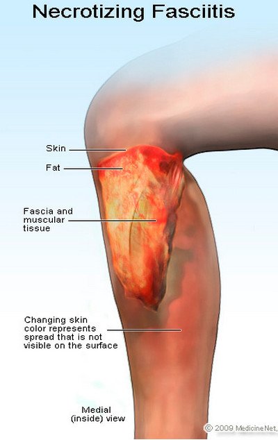
Necrotizing Fasciitis symptoms

Picture 1: Necrotizing Fasciitis Features

Image source : Medicine Net
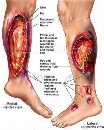
 

Necrotizing Fasciitis image

Picture 2: Necrotizing Fasciitis Picture

Image source : effguide

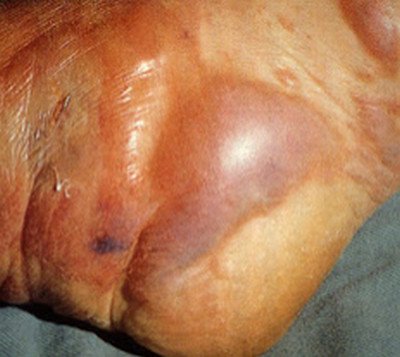
Necrotizing Fasciitis Picture
Picture 3: Necrotizing Fasciitis on leg (photo)
Image source : uga.edu

Necrotizing Fasciitis Diagnosis

There are several ways to diagnose necrotizing fasciitis.

  • A physician or medical practitioner would usually start assessment based on a patient’s symptoms as well as the precipitating and predisposing factors that resulted to the condition. An interview regarding past surgeries or even exposure to seawater may predispose a patient to necrotizing fasciitis.
  • In addition, a physical checkup of the affected area would affirm the physician’s suspicions regarding the disease.
  • Once established, the physician could confirm the possibility and existence of the condition through different blood tests as well as CT scans and skin biopsy.
  • Another more profound diagnosis tool would be through the use of the Laboratory Risk Indicator for Necrotizing Fasciitis (LRINEC) score. It uses a number of diagnostic tools to establish a positive result for necrotizing fasciitis. Greater than normal values for C-reactive protein, white blood cell count, creatinine, hemoglobin, and glucose plus an additional lower valuefor sodium would affirm a positive score for necrotizing fasciitis.

Treatment

  • Initial treatment for the condition would usually involve a broad spectrum set of antibiotics as waiting for a confirmatory diagnosis would delay treatment and possibly increase the risk of morbidity and mortality.
  • However, the preferred mode of treatment would still be surgical intervention. Patients with necrotizing fasciitis would require debridement 24 to 36 hours after the initial debridement and concurrently after so as to ensure complete cleansing of the entire wound takes place.
  • Once the causative factor is identified through Gram staining, appropriate antimicrobial therapy could be initiated. For Gram positive cocci, penicillin, clindamycin and ampicillin sulbactam are the drugs of choice. Meanwhile, piperacillin-tazobactam, imipenem-cilastatin, meropenem, ertapenem, cefotazime are the preferred drugs for Gram negative microorganisms. Ampicillin sulbactam could also be utilized for Gram negative bacteria.

Note that any kind of treatment should be done by a medical professional.

Prognosis and Possible Complications

Necrotizing fasciitis if left untreated or treated wrongly could have a poor prognosis. Although a rare condition, it can progress quite fast. Diagnosis and treatment should therefore be prompt to give the affected person a better chance. Necrotizing fasciitis could result to the admission of a patient to an intensive care unit.

Death could be the worst complication brought about by the condition. Other complications, which could also result, include the loss of an organ and tissue damage through debridement and sometimes, even through amputation.

It is therefore important that once necrotizing fasciitis is suspected, it should be promptly referred to a physician so that immediate treatment may be given. The delay in treatment of the condition could turn out a fatal and disastrous outcome for the patient.

References:

http://en.wikipedia.org/wiki/Necrotizing_fasciitis

http://www.medicinenet.com/necrotizing_fasciitis/article.htm

http://www.webmd.com/a-to-z-guides/necrotizing-fasciitis-flesh-eating-bacteria-topic-overview
http://emedicine.medscape.com/article/2051157-overview

Necrotizing Fasciitis

No responses yet

Osteoporosis

Dec 27 2012 Published by under Diseases and Conditions

Osteoporosis and its Classification

Osteoporosis is a condition characterized by increased porosity of the bones which can eventually lead to fractures. Osteoporosis is derived from the Greek word “ostoun,” which means bone, and poros meaning pore. It is characterized by a decrease in bone mineral density (BMD) and is most prevalent among women aged 35 and above. Osteoporosis is classified as primary type 1, primary type 2 and secondary osteoporosis.

Women suffering from this condition after their menopausal period belong to the primary type 1, which is also known as postmenopausal osteoporosis. Males and females who are above 75 years old would have the second classification, which is primary type 2 or senile osteoporosis. The last classification, secondary osteoporosis, may affect both males and females and may occur at any age. This usually results as a secondary effect resulting from a previous condition like a disease or excessive use of certain medications.

Who are at risk?

Risk factors for osteoporosis can be classified into nonmodifiable or uncontrollable risk factors and the modifiable or controllable risk factors.

Nonmodifiable factors would include the sexual orientation of an individual, menopause, the age, family history, ethnicity and those having a small stature.

  • Osteoporosis can affect both men and women. The latter, however, is more susceptible to developing osteoporosis especially after their menopausal stage. This is associated with the decreased levels of estrogen among women after menopause. Men, on the other hand, would have a decrease in their testosterone levels but the effect is usually not as profound as that of women.
  • Those who have family history of osteoporosis are also at an increased risk of 25 to 80% because of the genetic makeup of their bone mineral density.
  • Ethnicity and people with small statures are also at risk, examples of which are Asian women.

Modifiable factors would encompass alcoholism, tobacco smoking, calcium and vitamin D intake, malnutrition and exposure to heavy metals. In addition, certain disease condition increases the likelihood for osteoporosis. These would include immobilization, endocrine disorders like Cushing’s syndrome, malnutrition, renal insufficiency and those who are suffering from scoliosis. Women, who are pregnant or under lactation, are also at an increased risk. These factors can still be changed or adjusted.

  • Alcoholism, for instance, could be avoided as excessive alcohol intake increases the risks for bone fractures.
  • Smoking increases the breakdown of estrogen as well as predisposes a woman to an earlier menopause.
  • Decreased vitamin D and calcium intake is also a risk factor as these vitamins and minerals are necessary for bone strengthening.
  • Nutrition is also vital to ensure proper bone development and maintenance.
  • Exposure to heavy metals like cadmium and lead has also been linked to osteoporosis and osteomalacia, which is the softening of the bone.
  • Some disease and disorders have been associated with the development of osteoporosis. Immobilization, for example, could lead to osteoporosis especially if prolonged.  People with endocrine disorders like Cushing’s syndrome, hyperparathyroidism and hyoparathyroidism, thyrotoxicosis, diabetes mellitus type 1 and 2 as well as adrenal insufficiency are at greatest risk as well because of the role of regulation the endocrine organs play in the levels of calcium and phosphorus in the body.

Signs and Symptoms

  • Osteoporosis is actually a silent killer as it may not present with any symptoms for decades.
  • Pain and fracture are usually the first indication that a person may be suffering from osteoporosis. Fractures are usually caused by trauma or falls that a patient may be even unaware of.
  • The “dowager-hump” is probably the most profound symptom that is visible especially among older women. This is brought about by the chronic and repeated spinal fracture, causing the vertebrae to collapse and form a curved shape. The dowager hump also causes the chronic lower back pain experienced by the elderly.

Osteoporosis Diagnosis

Doctors would usually begin by having a look at the medical history of the patient. Risk factors may need to be identified so as to identify the underlying cause of osteoporosis. Mainly, osteoporosis can be diagnosed through radiography and by measuring the bone mineral density. Blood tests would also be included to measure calcium, vitamin D, phosphorus, testosterone levels as well as thyroid and kidney functions.

Bone mineral density tests would usually utilize the use of a specific radiographic examination. This is known as a dual x-ray absorptiometry scan (DXA). This is used diagnose osteoporosis by determining the bone density in the in specific areas. The test is usually precise and quite convenient to be done. Radiation exposure is also very minimal.

Treatment for Osteoporosis

  • Osteoporosis is best treated by a physician. Self-medication and self-treatment would not be able to resolve the condition other than prolong the appropriate treatment required in treating the condition.
  • Once signs and symptoms are noted or risk factors are identified, it would be best to have a physical examination done by a physician so that osteoporosis may be treated at the earliest possible time and prevent further consequences.
  • Treatment for osteoporosis would focus on slowing down the progress of the condition, improving bone density, controlling pain and preventing fractures and other complications. Treatment would include modification of diet and lifestyle, referral to a bone specialist as well as physical and occupational therapists and certain medications.
  • Estrogen replacement therapy remains to be the best option for women after menopause as it helps in slowing down bone loss. However, this is best done under the supervision of a physician as there are still conflicts surrounding estrogen intake among women after their menopause. Increased levels of estrogen have also been associated to coronary heart disease, stroke and thromboembolism. It is therefore important to have the proper dosing for estrogen replacement therapy.

Ways to Prevent Osteoporosis

The best approach against osteoporosis is to avoid it at the earliest possible time. This can be done by building strong bones through eating a well-balanced diet rich in calcium and vitamin D, regular exercise as well as avoidance of alcohol and smoking. Certain vitamins and mineral supplements can also help in the prevention of osteoporosis.

References:

http://www.medicinenet.com/osteoporosis/article.htm

http://www.nlm.nih.gov/medlineplus/osteoporosis.html

http://www.emedicinehealth.com/osteoporosis/article_em.htm


No responses yet

Pinguecula

Dec 26 2012 Published by under Diseases and Conditions

What is Pinguecula?

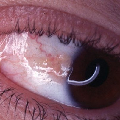
A pinguecula is a yellowish deposit on the conjunctiva adjacent to the cornea. It is a non-cancerous growth that results from calcium, fat or protein deposits. A pinguecula is almost similar to a pterygium, the latter, however, is wedged shaped. Although non-cancerous, it may grow larger over the years. A pinguecula would usually develop on the eye side closest to the nose. It is derived from the Latin word, “pinguis,” which means fat.

Causes of Pinguecula

  • Although the cause is still unknown, the most prevalent contributing factor for both pinguecula and pterygium is an exposure to ultraviolet radiation.
  • Prolonged eye irritation may also advance its development.
  • Dry eyes and environmental factors like wind and dust could also be a possible culprit for pinguecula to develop.
  • Exposure to harmful chemicals and mechanical damage to the conjunctiva may also precipitate the formation of a pinguecula.
  • Persons living in tropical climates also appear to have a greater risk of developing this eye condition.
  • In addition, persons who are over 40 have a higher risk but a pinguecula can also be common among persons aged 20 to 30 who spend significant amount of time under the sun.

Symptoms of Pinguecula

A person who has pinguecula usually does not experience any symptom other than the development of a yellowish nodule in the eye. If, however, it becomes irritated, a person may experience a feeling of having something in the eye.

Redness and swelling of the eye may occur if irritation persists and eventually worsens. If this happens, pingueculitis may occur. A pinguecula may also increase in size over the years.

Pinguecula Picture

Pinguecula Picture

Diagnosis

  • An eye examination done by an ophthalmologist would confirm the diagnosis for pinguecula
  • A slit-lamp may be used to allow the physician to examine the cornea, iris, lens, and the space between the cornea and the iris. Abnormalities would then be easily noted as the eye would be viewed at various small sections.

Treatments for Pinguecula

Once a pinguecula is suspected, it would be best to be referred to a doctor. Although enlargement of a pinguecula is possible, it would usually not require any form of treatment during the benign stages. Artificial tears or lubricating drops may be used to ease discomfort, redness or eye irritation. Stronger prescription drops may also be used to halt further enlargement of the nodule. Anti-inflammatory drugs may also be used to address discomfort and inflammation.

Can a pinguecula be removed?

Once a pinguecula is fully developed, it may no longer respond to topical treatments.

  • An established pinguecula may be removed once the nodule enlarges and threatens the eye sight as well as cause possible vision loss.
  • Extreme discomfort may also prompt the patient to have the pinguecula removed. This can be done as an outpatient surgery by an eye specialist.
  • A pinguecula may also be removed for aesthetic and cosmetic purposes especially once the yellowish nodule becomes too obvious.
  • In addition, those who are using contact lenses may find it difficult to wear their lenses thereby requiring them to undergo surgery.

The surgical procedure is usually painless and could be done in less than an hour. Topical anesthetics are used to minimize sensation during the surgery. After the procedure, eye drops instillation may be required to help ease swelling and prevent complications. Eye patch may also be necessary. Work activities may be resumed after a day or two as long as it would not causes excess strains on the eye.

Preventive Measures

However, undergoing surgery does not mean that the reoccurrence of pinguecula in the future would no longer be possible. It is still, therefore, important to avoid excessive exposure to UV light. Sunglasses with UV protection and safety occupational glasses are usually appropriate to use when frequent exposure to ultraviolet light is unavoidable.

In addition, this eyewear could also provide protection against environmental elements like dust and wind. Moreover, the use of artificial tears could also be beneficial once eye dryness is felt, especially during humid and dry conditions.

Pinguecula is an eye condition that may not need immediate attention of an eye specialist. But it would still be best to have a physician check it once noted or if discomfort is already felt. Treatment should best be done under a physician’s supervision more than self-medication and treatments.

References:

http://www.nlm.nih.gov/medlineplus/ency/article/001025.htm

http://www.umm.edu/ency/article/001025trt.htm


No responses yet

Hirsutism

Dec 25 2012 Published by under Women's Health

What is Hirsutism

Hirsutism is a condition common among women where excessive hair growth occurs in areas of the body in which terminal hair does not usually appear.

  • Hair growth among these women usually follows a male pattern of body hair – for example the growth of hair in the face, beard or chest area.
  • The condition may vary greatly among women.
  • Hair growth is usually fine and pale but may also grow coarse.
  • Hirsutism usually appears as a manifestation of an underlying disease condition especially if it develops after puberty has occurred.
  • Women are generally the ones affected by this condition. These women would usually tend to have increased levels of testosterone and other androgen hormones.
  • Androgen is the steroid hormone that stimulates male characteristics in an individual. An elevated level of androgen predisposes women to have excessive hair growth.

Causes of Hirsutism

The male hormone, androgen, is usually the culprit behind hirsutism. There are two course of actions in which androgen can cause hirsutism. The first one takes place when androgen levels are extremely elevated. On the other hand, oversensitivity of the hair follicles to androgen is the second factor that could lead to the condition.

Excessive production of androgen would increase hair follicle size, diameter and would usually quicken the hair growth phase. Hair and skin oiliness may also occur as a result of overproduction of androgen secretion. Also despite the excessive hair growth, hair loss in the scalp area occurs as overproduction of hair only takes place in androgen-sensitive regions like the sideburns in the face.

Certain conditions that may elevate a woman’s androgen levels include polycystic ovary syndrome, congenital adrenal hyperplasia, Cushing’s syndrome, acromegaly, ovarian tumors, certain medications and even obesity.

Signs and Symptoms of Hirsutism

  • Excessive hair growth in certain areas of woman’s face and body is the most obvious sign of hirsutism. It also usually follows male hair growth patterns. This means that hair growth usually takes place in areas common for hair growth among males like the beard, chest, face, and the areola. More advanced hirsutism would have hair growth on the upper back, sternum, the upper abdomen and on the shoulders.
  • Other telltale signs that a woman may be having hirsutism are when menstrual periods become irregular, acne attacks occur, the loss of feminine body shapes and usual secondary male characteristics like deepening of the voice, enlargement of the clitoris and broadening of the shoulders. These signs would usually be evident if hirsutism is primarily caused by hormonal imbalances.
  • However, if hirsutism is caused by an underlying medical disorder like Cushing’s syndrome, clinical manifestations may be different from a hormonal case. These signs and symptoms would include obesity, hypertension or elevated blood pressure and diabetes.

Who are at Risk for Hirsutism

The genetic makeup of an individual usually plays a role in inheriting hirsutism. South Asian and South and Middle European women are the races which have the most prevalent cases of hirsutism. The condition usually arises at the start of puberty and may continue to develop on if no proper treatment is undertaken.

How is Hirsutism Diagnosed

  • Physical assessment and a medical history are necessary to be able to diagnose and confirm hirsutism.
  • Menstrual cycles may need to be identified. Normal menstrual cycle patterns would usually indicate that the condition may be genetic. However, an irregular menstrual cycle would signify polycystic ovarian syndrome or some other underlying cause.
  • Blood workups may be required by the physician as well as other diagnostic tools like pelvic ultrasound, magnetic resonance imaging (MRI) and computed tomography (CT) scan. Blood tests would usually show elevated levels of androgen and testosterone. Meanwhile, ultrasound and other imaging studies would be necessary to identify cysts or tumors in the ovaries or adrenal glands.

Treatment for Hirsutism

Hirsutism is best managed by a physician. No amount of self-treatment or self-medication would relieve a person suffering from this condition. Women would usually find various ways of hair removal techniques to address this problem. But these treatments would not be addressing the real issue as the problem is not external but rather internal.

  • The use of Flutamide for a period of one year has been the most effective form of treatment among those affected by hirsutism. This should be done under the supervision of a doctor. For obese women, losing weight is vital as it will help in reducing androgen levels.
  • Home remedies for hirsutism would include drinking a cup of spearmint tea daily for 5 days. This has proven significant for women who had polycystic ovary syndrome as the underlying cause for hirsutism.
  • Surgical removal of tumors in the adrenals and ovaries will also be necessary if these would be the cause of hirsutism. In addition, psychological treatment may also be necessary especially for those experiencing frustration, embarrassment and low self-esteem.

Remedies for Hirsutism

  • Epilation or depilation can address the immediate problem of removing unwanted hairs. Shaving or trimming is probably the most common and convenient way of removing the excess hair. Chemical and commercially prepared depilatories are also available over the counter but they may irritate the skin. Waxing, sugaring and threading are also other ways to remove unwanted hair.
  • Laser hair removal could be another option but this could be impractical as not only is it expensive but hair growth would start appearing not long after the procedure as this does not address the root cause of hirsutism. Skin irritation could also result from this treatment so it would be best to seek a physician’s advice before going through with this procedure.

Hirsutism is a condition that is affecting a number of women. It used to be a stigma back in the days when excessive growth hair became unmanageable. But today hirsutism is no longer a problem and can be readily treated and managed under a physician’s direction. In addition, even if hirsutism is beyond a woman’s control, it is still necessary to avoid exposures to factors that may aggravate the condition.

References

http://www.umm.edu/altmed/articles/hirsutism-000081.htm

http://emedicine.medscape.com/article/1072031-overview

No responses yet

Ebola Hemorrhagic Fever

Dec 24 2012 Published by under Diseases and Conditions

What is Ebola Hemorrhagic Fever

Ebola virus disease (EVD) or Ebola hemorrhagic fever (EHF) is a severe fatal disease that can affect both humans and primates. Four viruses which causes the disease in humans are the Bundibugyo virus (BDBV), Sudan Virus (SUDV), Ebola Virus (EBOV) and Taj Forest Virus (TAFV), which is also known as the Côte d’Ivoire Ebola Virus (CIEBOV).

The disease got its name from the Ebola River in the Democratic Republic of Congo (formerly known as Zaire) in Africa where it was first recognized. The fifth virus, the Ebola-Reston, caused the disease among primates.This virus has recently been found in the Philippines.

Ebola hemorrhagic fever first broke out in Sudan and Zaire in 1976 causing one of the highest fatality rates at 90%.

How is the Ebola Virus Transmitted?

The Ebola virus belongs to the family Filoviridae. The disease can be transmitted to humans from infected animals as well as through direct contact by a human to infected body fluids or needles.

Nosocomial transmission during Ebola outbreak can also be possible. Although airborne transmissions have been documented among monkeys, there is still no known evidence of such transmission occurring among humans.

Signs and Symptoms

The signs are symptoms of quite similar to that of Marburg Virus Disease (MVD), which is also another type of viral hemorrhagic fever. The incubation period for the virus would usually take place between 2 to 25 days.

  • Fever, headache, muscle and joint pains, sore threat, and weakness are usually the first signs of Ebola hemorrhagic fever. These influenza-like symptoms may also accompanied by diarrhea and vomiting as well as stomach aches.
  • Sore throat, cough, dyspnea, hiccups and pharyngitis may also manifest as part of the illness.
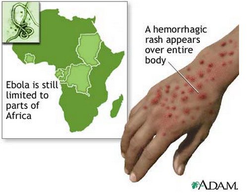
  • Hemorrhage is a late symptom of the disease. This could include bleeding from the eyes, ears and nose. In addition, eye swelling could also occur.Some people even experience genital swelling in their labia or scrotal area. Hemorrhage usually occurs as the virus tends to affect the ability of the blood to coagulate thereby causing bleeding in various parts of the body. Maculopapular rashes can also appear and be a form of bleeding in the skin. Hematemesis (vomiting of blood), hemoptysis (coughing up of blood) and melena (black, tarry stools) are also manifestations of possible bleeding with Ebola hemorrhagic fever.

The development of hemorrhage can be indicative of a poor prognosis as it can eventually lead to multiple organ dysfunction syndromes, focal tissue necrosis or disseminated intravascular coagulation.

Ebola Virus Pictures

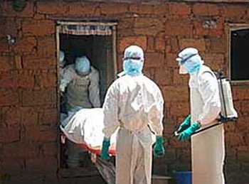
Medical Experts Handling blood from a patients with this condition is an extreme bio-hazard

Ebola Hemorrhagic Fever rash on hand

Picture 2 : Ebola Hemorrhagic Fever rash on hand

Image source : ADAM

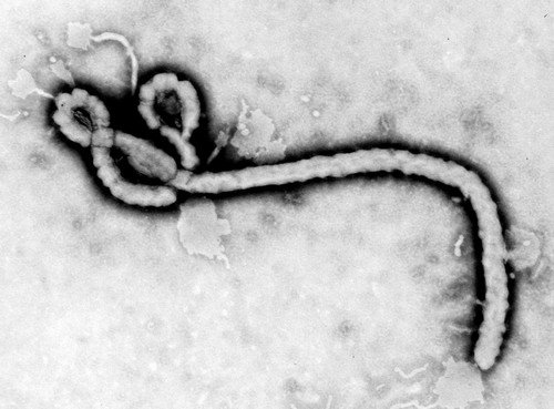
Ebola Hemorrhagic Fever Virus

Picture : Ebola Virus

Diagnosis of Ebola Hemorrhagic Fever

As Ebola hemorrhagic fever manifests similar signs and symptoms to other viral hemorrhagic diseases, it may be difficult to diagnose at the initial onset of the disease. Signs like red eyes and skin rashes could be indicative of various viral diseases. However, with proper physical assessment as well as interrogation of possible exposures to an Ebola virus, a definitive diagnosis may be made.

Taking a look into a person’s medical history would be an important tool in diagnosing the illness as the medical history would reveal a travel or exposure to wildlife. Confirmatory diagnosis for the Ebola hemorrhagic fever can be done through the isolation of the virus or through identification of the Ebola virus antigen or genomic or subgenomic RNAs in an infected person’s blood samples during the acute phase. Meanwhile, IgM and IgG antibodies could be tested during the later phase of the disease.

Treatment for Ebola Hemorrhagic Fever

Unfortunately, there is no specific treatment for Ebola hemorrhagic fever. Patients suffering from this disease would usually have supportive therapy. This would include minimizing invasive procedures, balancing fluids and electrolytes, anticoagulant therapy to prevent the occurrence of bleeding, oxygen maintenance, management of pain as well as the administration of antibiotics to address secondary infections.

A patient diagnosed with Ebola hemorrhagic fever would usually require an admission to the intensive care unit for better monitoring. Blood or platelet transfusion may also be necessary to correct coagulation problems and blood loss.

Ways to Prevent Ebola Hemorrhagic Fever

Prevention of Ebola hemorrhagic fever can prove a challenge as it is highly contagious and infectious. Personal protective equipment is necessary especially for those who are greatly exposed to the Ebola virus like medical practitioners and laboratory technicians.

Ebola hemorrhagic fever is a fatal condition that requires the immediate attention of a physician. Although no definitive treatments are available at this point, the worsening of the condition can be avoided and prevented as long as prompt medical treatment is given to a patient suffering from this condition.

References:

http://www.cdc.gov/ncidod/dvrd/spb/mnpages/dispages/Fact_Sheets/Ebola_Fact_Booklet.pdf

http://www.nlm.nih.gov/medlineplus/ency/article/001339.htm

No responses yet

Gangrene

Dec 23 2012 Published by under Diseases and Conditions

Gangrene Definition

Gangrene is the necrosis or death of a body tissue. It is the result of the loss of blood supply to a tissue. Tissue injury and bacterial invasion can also lead to gangrene. People having chronic health problems affecting blood circulation are usually the ones at greatest risk for gangrene. Gangrene is derived from the Greek term “gangraina” meaning putrefaction.

Causes of Gangrene

  • Infection or ischemia brings about the development of gangrene. This can be brought about by thrombosis or clots in the blood vessels. Conditions which results to decreased blood supply like peripheral vascular diseases, diabetes and chronic smoking increases the likelihood for ischemia leading to gangrene.
  • Infection can be caused by bacteria like Clostridium perfringens and Bacillus fusiformis.
  • The lower extremities are usually the most affected body part because it is the most distal for the distribution of oxygen through the circulating blood supply.

Classification of Gangrene

  • The most common classifications are the wet gangrene and the dry gangrene. Wet or moist gangrene is the most dangerous among the two. This occurs in moist tissues and organs like the oral mucous membrane, bowel, lungs, vulva and cervix. If left untreated, wet gangrene usually develops into sepsis and could quickly result to death in a matter of a few hours or days because of its poor prognosis. Wet gangrene develops mainly from venous or arterial blockage. Rapid growth of bacteria would then take place.  Toxins would then be produced by the bacteria leading to their absorption. Once this occurs, septicemia could result and eventually lead to death. The affected part would usually produce oozing fluid or pus, thus the term “wet.”
  • The second type of gangrene is dry gangrene. It usually occurs at the distal part of a limb and is often the result of an ischemia. Arterial occlusion is the primary cause of this classification of gangrene and could lead to the eventual sloughing of the tissue once necrosis sets in. The progression for dry gangrene is slower compared to wet gangrene and could take as much as several months until such that blood supply is no longer viable to keep the tissue. Gas production is absent so infection, pus and feelings of wetness are unlikely. People having problems with circulation are those who are at the greatest risk for developing gangrene. These could include diabetics, people suffering from arteriosclerosis and chronic smokers.
  • Other classifications of gangrene are gas gangrene, necrotizing fasciitis and internal gangrene. Gas gangrene is a type of gangrene caused by bacteria releasing gases that infiltrates body tissues. This type of gangrene often leads to shock and toxemia and is usually treated as an emergency case. On the other hand, necrotizing fasciitis is an infection of the fascia, which is a fibrous connective tissue in the body. Internal gangrene is a type of gangrene that occurs internally. Various internal organs may be affected and symptoms of wet gangrene may be visible once the organ is exposed by the surgeon.

Gangrene Photos

gas gangrene

Picture 1 : Gas Gangrene

wet gangrene

Picture 2  : Wet Gangrene

Dry gangrene

Picture  3 : Dry Gangrene

Symptoms of Gangrene

The symptoms of gangrene may vary according to the gangrene classification as well as the type of organ involved.

  • People with dry gangrene would usually experience numbness in the area and is cool to touch. Discoloration would usually start with redness to brown until it becomes black and shriveled. The area may appear mummified and could eventually slough off.
  • As for wet or moist gangrene, the area usually becomes swollen and is quite painful. Signs of decay like sloughed tissues, pus production and local oozing of fluid can take place. A foul odor may develop because of the toxins produced by the bacteria. Fever and other signs of sepsis may also develop.
  • In gas gangrene, the area usually becomes infected and bloody discharges are also produced just like in wet gangrene. Similar symptoms of wet and dry gangrene may also occur in gas gangrene. Internal gangrene, in contrast, has minimally obvious symptoms because of the location of the affected area. Usually, the skin overlying the area would become red, swollen and painful to touch. In addition, symptoms of wet gangrene are usually present.

Gangrene Treatment

Once a diagnosis of gangrene is established through physical assessment of the clinical symptoms, it is important that treatment would be given at the earliest possible time. Progression of the condition could be as fast as a few hours to a few days. It is therefore vital to seek medical attention once signs and symptoms of gangrene are noted.

The treatment options for gangrene would depend on the type of gangrene and the involvement of other body organs.

  • Debridement or the surgical removal of dead or injured tissue is the treatment of choice.
  • In worst scenarios, however, amputation may be necessary to prevent the progression of the gangrene to other uninvolved body parts.
  • Proper wound care is important as to prevent infection to settle in the new wound.
  • Antibiotic therapy is also important as a form of prophylaxis.
  • Psychological therapy may be necessary as feelings of low self-esteem and depression may be experienced especially for those having amputation.
  • Rehabilitation therapy is also another aspect which may be necessary to help the patient cope and re-adjust to the life without the amputated body part.

In addition, lifestyle modification may also be necessary especially for those who are chronic alcoholics and smokers. Diabetics should also have better control with their blood sugars. Moreover, education regarding proper wound care and prevention is possibly the best weapon against the recurrence of gangrene. Early identification of the signs and symptoms would enable a patient to seek treatment early.

Gangrene is therefore a medical condition that needs to be addressed by a physician. Self-treatment would not be possible for this condition and could in fact just worsen the situation as it would delay medical and surgical treatment. Gangrene therefore should be treated by a physician so as to prevent it from worsening as well as to be able to save the affected area at the earliest possible time.

References:

http://www.medicinenet.com/gangrene/page5.htm

http://www.emedicinehealth.com/gangrene/page11_em.htm#prevention


No responses yet

Craniosynostosis

Dec 22 2012 Published by under Diseases and Conditions

What is Craniosynostosis?

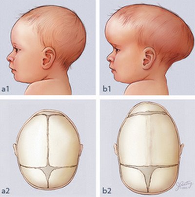
Craniosynostosis is an abnormal birth defect that causes the premature fusion of one or more sutures in a baby’s skull by ossification. This results in a distorted growth of the skull and an increase in the intracranial pressure. The fusion of the sutures would also cause the skull to develop parallel to the closed sutures instead of perpendicularly. This skull growth would give the appearance of an abnormal head shape and facial features.

 Craniosynostosis Types

Craniosynostosis is classified depending on the number of fusion of sutures involved.

  • Simple craniosynostosis refers to the involvement of a single suture only.
  • Complex or compound craniosynostosis refers to the premature fusion of more than one suture.
  • In addition, when craniosynostosis comes with deformities in other areas of the body like the limbs, heart, and central nervous system, it is termed as syndromic craniosynostosis.
  • Absence of other abnormalities would be referred to as non-syndromic or isolated craniosynostosis.

Causes of Craniosynostosis

The cause of craniosynostosis is still unknown. However, several researches have suggested that genetic mutation may play a role in the development of the condition. Several factors have been identified that poses a great risk for developing craniosynostosis. These factors usually occur during pregnancy. Some of these include womb constraint, maternal smoking and exposure to drugs containing amine. Increased levels of thyroid hormones have also been associated with craniosynostosis.

Craniosynostosis Diagnosis

Craniosynostosis is diagnosed through physical assessment, medical history and radiographic examination.

  • Physical assessment would show apparent signs and symptoms for craniosynostosis while medical history would check on the pregnancy condition of the mother as well as possible risk factors identified in both parents.
  • Radiographic analysis of the skull can be done through computed axial tomographic scan (CT scan) although plain radiographic examination may also be utilized. The main advantages, however, of a CT scan are the easy identification of the suture and structural abnormalities of the brain.

Signs and Symptoms of Craniosynostosis

The presenting signs and symptoms of craniosynostosis are usually dependent on the type of craniosynostosis a child has.

  • Generally, patients with craniosynostosis would present signs and symptoms of increased intracranial pressure (ICP), obstructive sleep apnea, skull deformities and neurobehavioral impairment.
  • Vomiting, vision problems possibly leading to vision loss and headache are usually the most common symptoms for elevated intracranial pressure. The increase in intracranial pressure is usually due to the continued brain growth in a rigid skull.
  • Abnormalities in the skull would include the absence of a fontanel and deformities in the shape of the head with no or slow physical growth. Physical assessment through measurement of head circumference is therefore important especially in cases with hydrocephalus and microcephaly. Physical deformities of the digits, neck, spine and toes may also be apparent especially among syndromal craniosynostosis.
  • Neurobehavioral impairment would include problems with attention, visual spatial skills, reading and language. Others may present with a decreased IQ but there are also other cases in which the child presents with normal intelligence.

Craniosynostosis Pictures

craniosynostosis of the lambdoid suture

Picture 1 : craniosynostosis of the lambdoid suture

Image source: wikipedia.org

craniosynostosis picture

Picture 2 : Craniosynostosis

craniosynostosis

Picture 3 : Craniosynostosis image

Treatment for Craniosynostosis

Surgical intervention is the best treatment modality for children with craniosynostosis. The goals for surgery is to relieve pressure on the brain, allow space for the brain’s growth and development as well as correct and improve the physical appearance of the child. Surgical intervention is usually done when the child is still an infant and could be a series of surgeries rather than just one surgical operation.

There are various conditions which need to be met prior to surgery.

  • The ideal age for surgery is between 3 to 6 months as surgery performed younger than 6 months are at an increased risk for relatively large losses of blood volume.
  • The presentation of how the sutures fused prematurely also plays an important factor on when to perform the surgery.

As craniosynostosis is a life threatening condition, it is best advised that a pediatric neurologist would evaluate the condition. Monitoring of the child’s condition should start from birth onwards until such time that surgical correction is performed. In addition, craniosynostosis would require a team of specialists to monitor and evaluate the child’s condition from physical health to the mental and behavioral health of the child.

A psychologist may be necessary as the child may experience feelings of low self-esteem and depression. Regular follow up consultation is also necessary to ensure a good outcome and prognosis for the child.

References:

http://www.ncbi.nlm.nih.gov/pubmedhealth/PMH0002557/

http://emedicine.medscape.com/article/1175957-treatment#showall


No responses yet

Sickle Cell Anemia

Dec 21 2012 Published by under Diseases and Conditions

What is Sickle Cell Anemia  ?

Sickle cell anemia is a severe, chronic, autosomal genetic blood disorder characterized by abnormal red blood cells. Normally, the red blood cells contain an iron-rich protein known as hemoglobin, which is responsible for carrying oxygen to the different cells in the body.

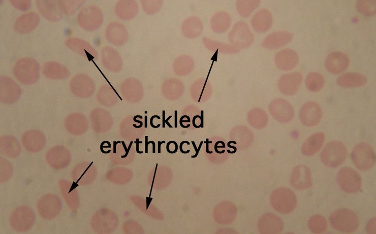
However, in sickle cell anemia, the abnormal hemoglobin is distorted and resembles rigid and sickle shaped or crescent-liked erythrocytes. Fragility among the red blood cells is increased but flexibility is decreased thereby resulting in various complications. In addition, these cells tend to clog the blood flow of blood vessels causing pain and organ damage. This is also called a sickle cell crisis or a vaso-occlusive crisis and mostly occurs in children.

Causes of Sickle Cell Anemia

Sickle anemia is caused by a genetic mutation of hemoglobin S. This condition is most prevalent among inhabitants of tropical and subtropical regions. Hemoglobin S is usually characterized by fragile, sickle shaped red blood cells which could lead to poor delivery of oxygen to the different tissues in the body.

In addition, the shape of the red blood cells enables it to become easily stuck in small blood vessels thereby interrupting the blood flow. It would take two parents who are both positive for the sickle cell trait to have their offspring test positive for sickle cell anemia. Meanwhile, individuals with the sickle cell trait do not usually have any symptoms and could have normal life expectancies.

Signs and Symptoms of Sickle Cell Anemia

  • Do not usually appear until after 4 months old
  • Characterized by a sickle cell crisis
  • Result of vaso-occlusive crisis, aplastic crisis, splenic-sequestration crisis and hemolytic crisis
  • In vaso-occlusive crisis, there is a blockage or obstruction in the blood flow due to misshapen Hemoglobin S. Patients may experience organ ischemia, pain and eventually necrosis. Meanwhile, aplastic crisis indicates worsening of anemia. Tachycardia, pallor and fatigue may be the usual symptoms. In splenic-sequestration, the spleen is usually the organ which is affected most resulting in the enlargement of the organ. This type of crisis usually requires emergency treatment because it is a high risk for circulatory failure. Lastly, hemolytic crisis occurs when the red blood cells breakdown faster than normal. This condition has been greatly associated with G6PD or glucose-phosphate deficiency.
  • Generally, the symptoms experienced by the patient once anemia becomes severe are shortness of breath, fatigue, pallor, and increased heart rate as well as a yellow or sallow discoloration of the skin. Pain experienced could be severe and may require hospitalization. The bones of the back, chest and long bones are usually the most sensitive body parts. Some children may also experience abdominal pain.
  • Once the organs are already affected, specific symptoms may already become apparent depending on the organs involved. Visual loss, painful and prolonged erection, confusion and ulcers are some of these symptoms.
  • Bone infection, pneumonia, cholecystitis or the inflammation of the gallbladder could also result as complications especially after the spleen becomes damaged. Arthritis, delayed growth and puberty are also possible.

Diagnosis

  • A blood smear test may be done to detect and quantify the types of hemoglobin present.
  • A hemoglobin electrophoresis test may be required to confirm the type of hemoglobin in the blood.
  • Other diagnostic tests that may also be necessary are complete blood count, blood oxygen, bilirubin, serum creatinine and potassium as well as sickle cell test.

Sickle Cell Anemia histology picture

sicke cell anemia histology

picture : sicked erythrocytes

image source  : cord.edu

Treatment for Sickle Cell Anemia

Unfortunately, there is no cure for the condition. Treatments are usually palliative and aim to improve anemia as well as prevent further damage and complications. Other goals would include pain management, prevention of infection and possible stroke.

  • Fatigue and anemia brought about by sickle cell anemia are usually managed with blood transfusions to restore hemoglobin within normal levels. Blood transfusions, however, has its risks and these include blood borne diseases like hepatitis, allergic reactions, infections and iron overload.
  • Pain management would include over the counter drug preparations if the pain is not too severe as well as plenty of fluids and adequate rest. However, pain may need to be managed in a hospital setting once it becomes too severe. Acetaminophen (Tylenol) or nonsteroidal anti-inflammatory drugs (NSAID) are the medications given for mild to moderate pain. Opioids would become the drug of choice for extremely severe pain. Adequate hydration is also necessary as well as oxygen therapy if the oxygen saturation becomes too low.
  • Hydroxyurea is a medication given to those who have sickle cell anemia. It prompts the body to produce fetal hemoglobin or Hemoglobin F, which is the type of hemoglobin newborns have. Hemoglobin F would tend to replace Hemoglobin S thereby eliminating the cause of sickle cell anemia. Hydroxyurea is also a medication used in chemotherapy.
  • Stem cell, blood and bone marrow transplants are also fast becoming popular as the preferred treatment for sickle cell anemia. However, research is still ongoing regarding these treatments but bone marrow transplants have been proven effective among children.
  • Sickle cell anemia is a condition that would require the management of a physician. Self-medication would not be applicable in this condition as it could only worsen the situation. In fact, sickle cell anemia would require the need of not just one specialist but several physicians as these could be a systemic disease affecting multiple organs. It is therefore important to have a physician manage the condition rather than rely on self-treatments and hearsays.

References:

http://www.nhlbi.nih.gov/health/health-topics/topics/sca/treatment.html

http://www.nhs.uk/conditions/Sickle-cell-anaemia/Pages/Introduction.aspx

http://www.medicinenet.com/sickle_cell/page3.htm

No responses yet

Vasculitis

Dec 20 2012 Published by under Diseases and Conditions

Vasculitis is a general term used to refer to a group of disorders that destroys the blood vessels through inflammation. Vasculitis may affect arteries, veins and the capillaries. It is primarily characterized by an inflammation and damage to the walls of the blood vessels. Vasculitis is derived from the Latin word “vasculum,” which means vessel, and “itis” meaning inflammation.

What Causes Vasculitis

Vasculitis may be classified according to its cause as primary vasculitis or secondary vasculitis.

  • In its many forms, the cause of primary vasculitis is unknown.
  • As for secondary vasculitis, there is usually an underlying cause of the condition. These could include infections, immune system diseases, allergic reactions and blood cell cancers. In some conditions, vasculitis may be related to an ongoing infection as it tends to trigger an autoimmune response that targets the blood vessels. Hepatits B and Hepatitis C virus are two of the most common infection that can cause polyarteritis nodosa. On the other hand, immune system diseases that can lead to vasculitis include reheumatoid arthritis, scleroderma, lupus and Sjögren’s syndrome. Allergies and certain medications could also predispose a person to develop vasculitis. In addition, blood cell cancers like leukemia and lymphoma can also cause vasculitis.

Types of Vasculitis

Vasculitis may be classified according to its cause, the type of blood vessel, its location or its size. Phlebitis (veins) and arteritis (arteries) are the classification of vasculitis dependent on the type of blood vessel involved.

Signs and Symptoms of Vasculitis

Signs and symptoms of vasculitis vary greatly depending on the blood vessels involved.

  • General symptoms would include fever, weight loss and appetite loss.
  • The skin may also develop palpable purpura, which are red or purple discoloration of the skin brought about by bleeding in the tissues.
  • Nervous systems problems such as numbness, weakness, reduced visual acuity or vision loss, stroke, tinnitus and headache may also be experienced.
  • Other symptoms could also include nose bleed, bloody cough and stool as well as abdominal pains.

Diagnostic tools for Vasculitis

  • Blood tests and body fluid tests of a person suffering from vasculitis would usually indicate inflammation.
  • In addition, certain organ function tests would indicate abnormality. For instance, creatinine levels would be elevated if the kidneys are already affected.
  • Biopsy of the blood vessels would be the defining factor to confirm the diagnosis for vasculitis. This could include tissue samples from the skin, lungs, nerves, sinuses and kidneys.
  • Angiogram may also be indicated as an alternative to biopsy. It is a radiographic imaging of the blood vessel that would require the injection of a contrast medium. However, this procedure is contraindicated for people who may have dye allergy or IV allergies as this utilizes a radiopaque contrast medium.

Treatments for Vasculitis

There is no specific cure for vasculitis. Instead, the main goal of treatment is to stop the inflammation and to suppress the immune system to prevent further attacks on the blood vessels. Preventing serious complications is also an important treatment plan.

  • Corticosteroid medications like prednsisone are usually the treatment of choice as it suppresses the inflammatory response of the immune system.
  • Other immunosuppressive drugs like cyclophosphamide may also be considered depending on the case of severity as well as the type and location of a patient’s vasculitis. However, it is important to note that taking in immunosuppressive drugs should be done under the physician’s orders as these are usually corticosteroids. This means that these drugs are synthetic hormones that have a wide range of effect encompassing physiological processes like stress and immune response, carbohydrate metabolism, blood electrolyte levels, protein catabolism and inflammation. They should therefore be regulated to ensure proper dosing of the drug.
  • Moreover, drug treatments utilized are usually combined therapies so it is also important that a patient inform their physician regarding other drugs that the patient is also taking in.
  • Also, there are cases of vasculitis that would require the management of a multidisciplinary group. This is because some cases may need specialists to address specific organ problems like the heart and kidneys, which would require the attention of a cardiologist and nephrologist respectively.
  • Topical medications may also be used and applied to sores. These could help address the pain and discomfort as these usually contain anti-inflammatory agents like corticosteroids and even anesthetics in other preparations.

In addition, the symptoms of vasculitis may be controlled as long as the patient complies with proper medication, eat a well-balanced diet, practice a healthy lifestyle and exercise regularly. Swimming and walking are the best forms of exercises once the symptoms of vasculitis have disappeared. Another important thing to know is that remission may also be prevalent among patients suffering from vasculitis so it would be best to have regular check up with one’s physician to prevent its recurrence.

References:

http://www.nlm.nih.gov/medlineplus/vasculitis.html

http://www.mayoclinic.com/health/vasculitis/DS00513/DSECTION=treatments-and-drugs

http://emedicine.medscape.com/article/1008239-treatment

No responses yet

© 2023 Healthooze.com. All Rights Reserved. Privacy Policy