Archive for March, 2017

Pediculosis Capitis

Mar 14 2017 Published by under Diseases and Conditions

What is Head lice/ Pediculosis capitis?

A pediculosis capitis or head louse is a very contagious condition. The condition is very common, and you will most likely get it from day cares, nurseries or schools.

The condition is brought by the infestation of the famous head louse known a pediculus humanus capitis. It causes a lot of itchiness to the people who get it. [1,2]

Hair and scalp infected by lice and nits
Image 1: Hair and scalp infected by lice and nits
Picture Source: www.skinsight.com

Lifetime

Lice are tiny common insects that consume the human blood. The louse attaches its nits at the base of an individual’s hair close to the scalp. The eggs take up to 7 to 10 days before they can hatch. The adult louse isn’t able to live outside the head for over two days.

However, the nit can live for ten days away from the head. It can survive on the hairbrush, cloths or the carpet.  These lice can get from one person to the other by sharing items that have lice or head to head contact.[4,5,6]

Who can get the condition?

  • Head lice can get anyone. However, children are more prone to the condition, especially the ones aged three to eleven years. The socioeconomic group an individual belongs to doesn’t determine whether the person will get the condition or not.  Cleanliness of a person or even the environment doesn’t matter too.
  • The lice can quickly grasp some hair types better than others. People with smooth hair are more affected compared to the people with curly hair.  Females tend to get the condition more than the boys because of their long hair.
  • Head lice are common in places where people have close contact especially playgrounds, home, camp or schools. These insects consume human blood only, so someone cannot get the infection from pets or any other animal.[4,5,8]

Signs and Symptoms

For individuals with this contagious infection, non-moving eggs or moving lice will see on the hair or the scalp.  The size of the louse is 1-3mm, and they are whitish grey in color.  These lice do not fly or even jump. They just crawl, so it is easy to identify them.

  • The nits are small compared to the lice, and they are approximately 0.5 to 1 mm. They are white in color, and most of the time, they are firmly attached on the hair or near the scalp.
  • People with the condition can get some tiny red bumps or even sores on the neck, scalp or the shoulders.
  • Some people can also get swollen lymph nodes at the back of the ears and neck.
  • The lice can get to the eyelashes too, making the yes to look red or irritated.
  • When head lice infect someone, they cause a lot of itchiness, and they can make someone scratch the affected area a lot.  This can result in an infection or even scabbing.
  • The affected children can become very irritable and have some difficulties when sleeping.[8,9]

Self-care Tips

When you suspect that your baby has head lice, get a fine-toothed comb and try to look for the insects.  There is a particular louse comb in most drugstores nowadays, and finding these bugs will not be difficult.  Examine the baby careful using some bright light, and if possible, use a magnifying glass.

If you discover that your child has the louse of the nits, follow these tips to help get rid of them:

  • Several effective medications can be purchased over the counter to deal with the infection.  If you get the right medication, you will be able to kill the live lice. The eggs can prove to be difficult, but if you keep reapplying the drug, the newly hatched lice will be killed too. In seven or 10 days, the condition will be eliminated. Medication such as Pyrethrins or permethrin lotion can help. However, children who are below two years should use them. These medicines are absorbed into the skin, and this is not good for the young kids. These medications should be applied in very small quantities too.
  • It is advisable to avoid using a conditioner before applying over the counter medication. A conditioner will help coat the hair, preventing the insects from the drug. After you have used the medicine, do not wash the hair for a day or two.
  • After treating the lice, change into clean cloth. Wash all the beddings, the rest of the cloths and even towels using very hot water and use a hot cycle to dry them. This will ensure that all the lice in the cloths are killed.
  • If there are any objects, the child has come close to in 48 hours, wash them in very hot water for around 5 minutes.
  • If you have any contaminated objects that are not washable, seal them using plastic bags for two weeks, and this will ensure that the nits and lice are dead.
  • Ensure that you vacuum the floors well. The furniture too should be vacuumed.
  • Check everyone in the house and ensure everyone is free from the head lice. If anyone is infected, treat them too.
  • If your child is going to school, talk to the teacher, nurse or the caretaker about the child’s condition.
  • It is not wise to share a comp, hat, toy, hairbrush, beddings, cloths, or any contaminated items with an individual who has head lice. [5,6,8]
    Home fumigation is not required.

When should someone seek medical attention?

Before starting to treat your child at home, it is important to talk to your doctor.  Have any questions; do not do the treatment at home. The doctor should recommend the best medication to use.

  • When you notice any symptoms of a bacterial infection on the child’s scalp, call the doctor immediately. Bacterial infection can cause swelling, pus, pain and redness on the scalp.
  • For any pregnant mother, it is advisable to seek medical attention before applying the lice medication.

It is very important to ensure that you consult the doctor to confirm the presence of the head lice and the type of medication to use. [9,10]

References :

  1. Symptomview.com. The Bodyguard of your health – SymptomView.com [Internet]. 2016 [cited 20 January 2016]. Available from: http://www.Symptomview.com
  2. Pharmapacks.com. Pharmapacks – Health & Beauty Marketplace [Internet]. 2016 [cited 20 January 2016]. Available from: http://www.pharmapacks.com
  3. Emedicine.medscape.com. Pediculosis and Pthiriasis (Lice Infestation) Treatment & Management: Approach Considerations, Pesticides, Occlusive and Nonpesticide Therapy [Internet]. 2016 [cited 20 January 2016]. Available from: http://emedicine.medscape.com/article/225013-treatment
  4. [Internet]. 2016 [cited 20 January 2016]. Available from: http://www.skinsight.com/…d/pediculosisCapitisHeadLice.htm
  5. Uptodate.com. Pediculosis capitis [Internet]. 2016 [cited 20 January 2016]. Available from: http://www.uptodate.com/contents/pediculosis-capitis
  6. Sayler K. Pediculosis and Scabies: A Treatment Update – American Family Physician [Internet]. Aafp.org. 2016 [cited 20 January 2016]. Available from: http://www.aafp.org/afp/2012/0915/p535.html
  7. H F. Pediculosis capitis: new insights into epidemiology, diagnosis and treatment.  – PubMed – NCBI [Internet]. Ncbi.nlm.nih.gov. 2016 [cited 20 January 2016]. Available from: http://www.ncbi.nlm.nih.gov/pubmed/22382818
  8. [Internet]. 2016 [cited 20 January 2016]. Available from: http://www.mayoclinic.org/…head-lice/basics/definition/..
  9. [Internet]. 2016 [cited 20 January 2016]. Available from: http://www.skin-disorders.net/…es/pediculosis-capitis.html
  10. Schweinitz D. Pediculosis and Scabies – American Family Physician [Internet]. Aafp.org. 2016 [cited 20 January 2016]. Available from: http://www.aafp.org/afp/2004/0115/p341.html
No responses yet

Erythroplakia

Mar 12 2017 Published by under Diseases and Conditions

What is Erythroplakia?

Erythroplakia refers to an oral condition that appears as red lesions on the mucus membrane but cannot be attributed to any other illness or condition [1].

The reddish color of the patches has been attributed to lack of a keratin layer on the mucosal tissue. Instead of the keratin, there is a papillae connective tissue with enlarged capillaries. The capillaries happen to be in close proximity to the surface and that also plays a role in the reddish color [2].

The condition is similar to leukoplakia, which is characterized by white spots on the mucous membrane of the mouth [1, 3].

The condition is prevalent among men between the age of 60 and 70 years [2]. Together with leukoplakia, erythroplakia is considered to be a premalignant lesion [4]. As such, timely diagnosis and treatment is of great important to avoid development of cancer in the lesions. Routine oral check-ups will ensure the condition is detected early enough [5].

What does Erythroplakia look like (Pictures) ?

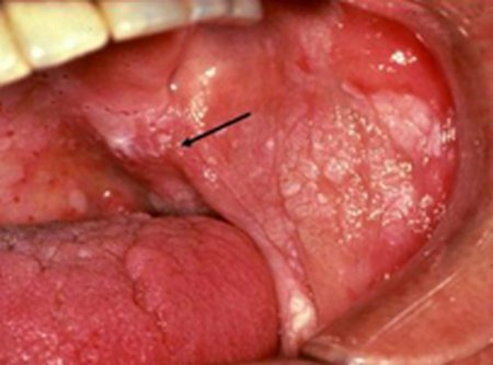
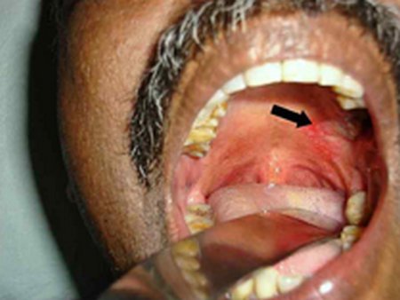
Erythroplakia lesions on the palate

Image 1: Erythroplakia lesions on the palate (Indicated by the arrow)
Photo Source: screening.iarc.fr


Erythroplakia on the tongue

Image 2: Erythroplakia on the tongue
Picture Source: screening.iarc.fr


History of Erythroplakia

  • A non-hemorrhagic, painless, red color lesion was observed on the upper part of the gut. This was observed and reported in 1852 [2].
  • Nevertheless, the first medical description of the condition in an academic paper was done in the year 1911 by Queyrat. In the paper, Queryat described a red patch on the glans of the penis of a person with syphilis. Queyrat coined the term erythroplakia [6].
  • Rubin named incipient carcinoma with the aim of describing the erythroplakia microscopic properties he observed in the same era. The precancerous nature of the lesions has become the most widely used histo-pathologic diagnosis Erythroplakia [2].

Sings and Symptoms

  • Erythroplakia is asymptomatic [4].

Signs

  • Red patches on the mucosal and/or sub-mucosal surfaces
  • Occurrence of pain may be an indication that the lesions are entering the malignant phase [2].

Cause:

  • It is idiopathic thus may come up spontaneously without a known cause [2, 6].

Risk Factors include

  • Smoking tobacco
  • Excessive consumption of alcohol
  • An opportunistic infection or a candidiasis super-infection. This could be associated with the oral-mucosal surface cells. Candida albicans has been observed severally in the erythroleukoplakia patches. The white and/or the red components of the lesions may diminish after the patient has received antifungal therapy [3, 7].

Clinical Features of Erythroplakia

1.    Occurrence varies with:

  1. Age: mean age at diagnosis of erythroplakia is 50-69 years, with less than 5% of diagnoses patients being under 30 years of age. Older patients also have a higher the risk of for the lesions to undergo the premalignant to malignant transformation.
  2. Sex: Erythroplakia is predominant among men. However, recent ratios show an increase in female patients, possibly owing to an increase in female smokers [8].

2.    Sites:

  1. Generally, Erythroplakia can occur on any of the mucosal surfaces around the neck or head [2].
  2. However, up to half of the diagnosed cases have been noted to exist on vermilion and intraoral surfaces. The rest of the recorded occurrences are divided between larynx and the pharynx.
  3. Vermillion patches are quite common and are most often found on the lower lips.

3.    Homogenous form:

  1. Usually found on the buccal mucosa and the soft palate. Sometimes they are found on the tongue or the floor of the mouth, albeit rarely. [2].
  2. This form is occurs as a soft, red, and velvety lesion with straight margins that are well-demarcated.
  3. Can be quite large in size in terms of the area they cover.

4.    Size:

Usually less than 15mm in diameter. However, some lesions are more than 4cm in diameters.

5.    Margins:

The lesions contrast sharply with the normal pink mucosa surroundings in the mouth. [3, 7].

6.    Granular/Speckled form:

They comprise of red and soft lesions. They are raised slightly and have regular outlines. The surface is finely nodular and speckled tiny plaques which are white in color.

7.    Smooth erythroplakia:

A palpitation test reveals a soft texture which is commonly described as a velvety feel on the fingers. The pebbled patches/lesions are a little firm but the erythroplakia doesn’t become really indurated until an invasive carcinoma develops within [2].

8.    Erythroleukoplaka:

  1. This condition is commonly observed where erythroplakia appears adjacent to or in same area as a leukoplakia in the mouth [9].
  2. The red spots are most probable to contain or develop dysplastic cells. Therefore, the red areas have to be biopsied readily and clinically examined with uttermost care.
  3. Erythroleukoplaka patches are quite irregular and usually not as bright as the erythroplakia homogenous type. They are found on the floor part of the mouth and on the tongue in most occasions.
  4. The borders are not as clear as in the homogenous lesions and they may blend with the surrounding oral mucosa surface.

Erythroleukoplakia

Image 3: Erythroleukoplakia
Photo Source: medscape.com

9. Unlike the leukoplakia lesions, erythroplakia covers large areas in the mouth. In addition, it rarely expands laterally after the initial diagnosis. However, it might be an artifactual property because most of the lesions are destroyed or removed totally after diagnosis.

Diagnosis

  • Erythroplakia rarely covers large parts of the patient’s mouth. While examined by use of hand, the erythroplakia feels soft. It remains soft without hardening till a persistent carcinoma develops inside the lesion [10].
  • Although some of the people with the condition may experience a burning, or sore sensation, erythroplakia is asymptomatic. Oral erythroplakia possesses the greatest risk of attaining malignancy compared to the other types of premalignant lesions.
  • Medical examination of the diagnosed erythroplakia reveals severe dysplasia of the epithelium or invasive cancer [11]. All erythroplakia lesions should be examined for malignancy with extreme clinical caution and suspicion. This is because erythroplakia lesions are highly likely to contain the histological foci of chronic dysplasia, or even invasive cancer.
  • Excision biopsy is advised and proper histological examination should follow as a compulsory practice every time there is clinical diagnosis of erythroplakia.

Treatment and Management

  • Treatment involves biopsy of the lesion to identify extent of dysplasia [10].
  • Complete excision of the lesion is sometimes advised depending on the histopathology found in the biopsy. Even in these cases, recurrence of the erythroplakia is common and, thus, long-term monitoring is needed.
  • The management of the condition involves taking measures that avoid the transformation into malignancy [7]. Creating public awareness about the illness, closely monitoring those that have undergone treatment and regular oral check-ups are sure ways of controlling it [4, 8].

References:

  1. http://www.oralcancerfoundation.org/cdc/cdc_chapter4.php
  2. http://dentalproblems.ygoy.com/2011/10/18/oral-precancer-erythroplakia-overview
  3. http://emedicine.medscape.com/article/1840467
  4. http://www.ada.org.au/app_cmslib/media/lib/1108/m324364_v1_villa.pdf
  5. http://www.webmd.boots.com/cancer/cancer-of-the-mouth-and-throat-overview
  6. https://en.wikipedia.org/wiki/Erythroplakia
  7. http://screening.iarc.fr/atlasoral_list.php?cat=A3&lang=1
  8. http://www.cancer.ca/en/cancer-information/cancer-type/oral/oral-cancer/precancerous-conditions/?region=on
  9. http://www.kcl.ac.uk/dentistry/about/acad/oral-leukoplakia-and-erythroplakia.pdf
  10. http://www.rightdiagnosis.com/e/erythroplakia
  11. http://www.healthcommunities.com/head-and-neck-cancer

No responses yet

Sunspots on the Skin

Mar 10 2017 Published by under Diseases and Conditions

What are sunspots on skin

These are dark portions or spots of the skin. They are also called solar lentigines (1, 2).

They develop more often after exposure to direct sunlight (2). Sunlight has ultra violet rays (UV rays) which are responsible for the development of these spots hence the name solar Lentigines (1). Exposure to the sun darkens and increases the number of the spots (2)

What causes the Sunspots on Skin?

The sunspots on the skin are more frequently caused by prolonged exposure to UV light from the sun (1). They are also caused by exposure of UV lights from tanning beds (2).

When the skin is exposed to the UV rays, special cells of the skin called melanocytes produce increased amounts of a substance called melanin in that region (2). Melanin pigment usually protects the skin against the effects of the UV rays (3). Every time the skin is exposed to the sun (UV rays) melanin is synthesized and the skin appears darker (3, 4).

Some authors also refer to them as

  • Freckles (5),
  • Senile freckles or old age spots (6)
  • Liver spots (7).

They were called liver spots because initially, they were thought to all be caused by liver problems (8)

In an individual with sunspots on the skin, there is accumulation of melanin within the skin cells (keratinocytes) due to increased synthesis by the melanocytes (6).

Note that the number of melanocytes varies as distinguished by Hunter, Savin and Dahl (9), even if other authors have used the names interchangeably. The author notes that in contrast to freckles, lentigines have increased number of melanocytes.


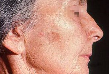
Sunspot on the side of the face
Image 1: Sunspot on the side of the face

Picture Source: emedicine.medscape.com

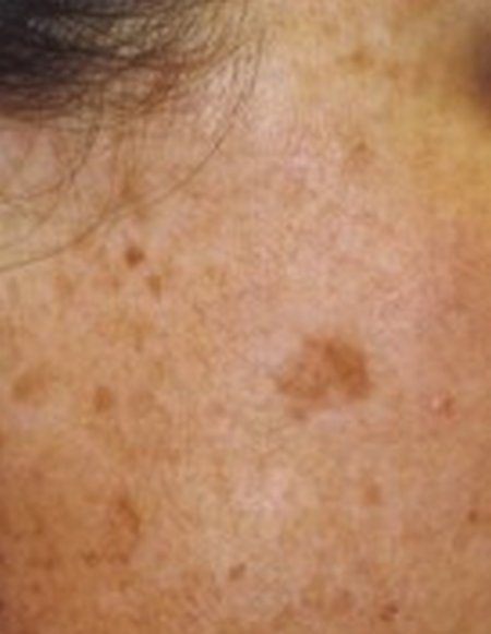
Sunspots on the side of the face

Picture 2: Sunspots on the side of the face
Photo Source: www.drbaileyskincare.com

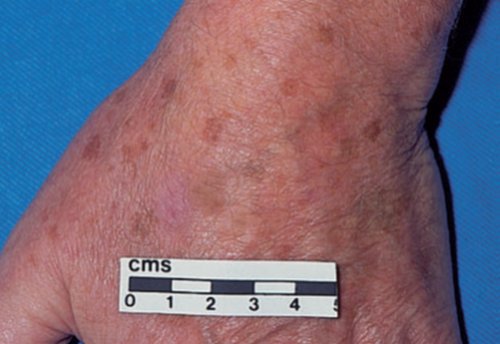
Sunspots on the back of hand

Image 3: Sunspots on the back of hand

Picture Source : Hunter, Savin and Dahl. Clinical Dermatology 3rd Edition. Massachusetts: Blackwell Science LTD 2002

Who can develop the Sunspots?

People of all ages are susceptible if they get exposed to UV rays without proper protection. However;

  • Those with lighter skin develop sun spots more frequently than those with darker skin (1, 7). This is because darker persons have better natural pigment protection of melanin as compared to light skinned persons (7, 10). This makes them more resistant to sun’s harmful effects(10)
  • Albinos have no melanin in their skin and therefore there are more at risk of developing sun spots and more vulnerable to the damaging effects of the UV rays
  • Older persons above 40 years will commonly have the senile freckles (6)

Where do you commonly develop Sunspots?

The Lentigos are usually brown to dark colored spots on the skin. Usually they will appear on the areas of great exposure to the UV rays. These areas include (2)

  1. Top of the scalp
  2. Sides of the face
  3. Sides of the neck
  4. Back of the hands
  5. Other areas exposed e.g back of the body if someone was sun bathing

How to get rid of Sunspots on skin ?

Sunspots are not dangerous but they cause discomfort since they are unsightly. However, it would be important to see a dermatologist to be sure that the dark /brown spots are actually sunspots because the spots of a melanoma (skin cancer) appears the same (2). Sunspots have been said to obscure the diagnosis of a melanoma. To distinguish the two, the doctor will perform a biopsy.

The sunspots spots can be eliminated via several methods (1). The following are suggested;

  • Application of creams, lotions and other cosmetic products to remove the spots
  • Eating certain foods to boost their levels in the body
  • Medical treatments

The products that can be applied on the skin and have been shown to fade the spots are;

  • Sun block creams – one should use a sunblock every time they anticipate exposure to the sun. It is recommended that sun screens should be worn everyday over exposed skin (4). It not only promotes healing of the skin but also protects against future formation of these spots. A minimum of SPF 15 must be used for optimal effectiveness(1)
  • Do IT yourself remedies – these are remedies that one does not have to purchase. They can easily be made in the comfort of the home. They are natural products that are also easily available. These include
    a.    Aloe Vera gel applied twice a day
    b.    Lemon juice
    c.    Fresh tomato
    d.    Apple cider vinegar and onion juice – mix equal parts of onion juice with apple cider vinegar. Apply the mixture on the skin for 10-5 minutes. Wash off with water
    e.    Garlic juice which can either be applied on the areas of the skin with the spots or can be drunk
    f.    Mixture of cod liver oils and castor oil in equal parts. Apply to the skin twice a day
  • To prevent sun spots it is important to avoid UV rays exposure by (4, 10)
    a.    Applying sunscreen at all times over areas exposed to the rays
    b.    Wearing hats
    c.    Seeking shade whenever possible to avoid direct sunlight if possible

What to include in diet to reduce Sunspots

Eating the following foods provide nutrients that help to reduce and eliminate sunspots;

  1. Fruits and vegetables Citrus fruits contain vitamin C that the body requires to repair worn out tissues. Eat plenty of oranges, tangerine and passion fruits. Watermelons, grapes and tomatoes contain a product called lycopene which help the body avoid sunburns. Green vegetables provide vitamins B and E essential for skin health (1)
  2. Green tea- green tea provides antioxidants (1)
  3. Fish


Medical and Surgical Treatments

Even though active treatment is not needed, topical creams may be prescribed to clear the spots on the skin. These may include skin lightening creams for example hydroquinone which suppresses melanocyte metabolic processes (7) and use of retinoid cream.

In surgical treatments, freezing with liquid nitrogen in cryotherapy has also been found to be effective for solitary spots (7). Chemical peels and Laser treatment may also be done.

References :

  1. MDHealth.com. Sunspot on skin http://www.md-health.com/Sunspots-On-Skin.html
  2. http://www.drbaileyskincare.com/blog/sunspots-on-skin/
  3. Waugh A and Grant A. Ross and Wilson anatomy and Physiology in health and Illness. 10th edition. Edinburgh: Elsevier; 2006
  4. http://www.drbaileyskincare.com/blog/skin-sun-spots/
  5. Freckle.  https://en.wikipedia.org/wiki/Freckle
  6. DermNet NZ. solar Lentigo http://www.dermnetnz.org/lesions/solar-lentigo.html
  7. Schwartz, R. A Lentigo. http://emedicine.medscape.com/article/1068503
  8. Liver spot. https://en.wikipedia.org/wiki/Liver_spot
  9. Hunter, Savin and Dahl. Clinical Dermatology 3rd Edition. Massachusetts: Blackwell Science LTD; 2002
  10. The Merck Manual of Medical information. Merck Research Laboratories; 1997

No responses yet

Herpetic Whitlow

Mar 08 2017 Published by under Diseases and Conditions

What is Herpetic Whitlow?

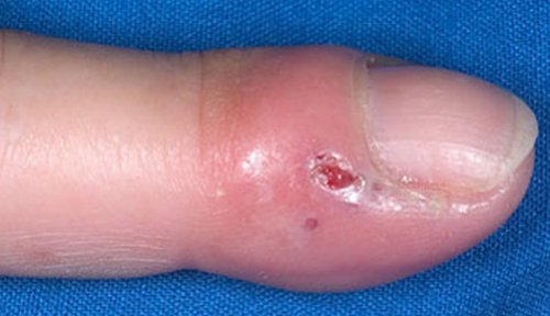
This is a laceration that appears on the thumb caused by the herpes simplex virus HSV-1 or HSV -2. Herpes originates from a Greek word meaning “to creep”.

This explains one characteristic of all herpes since they have a tendency to move along from one nerve end to the other. They may remain inactive in the body for quite some time before they replicate and affect the patient.  Everyone can be affected by this disease.

There are two types of Herpetic Whitlow.

  1. Herpes simplex virus- type 1    (HSV-1)
  2. Herpes simplex virus- type 2    (HSV-2)

HVS-1 is mostly contracted by medical workers who may have accidentally acquired it in their line of duty.  HSV-1 is the main causative agent of herpetic whitlow with approximately 60% cases and the rest of the 405 covered by HSV-2.

HSV-1 mostly affects fingertips, mouth and lips. This lesion usually affects the fleshy part of the finger tip. Patients with HSV-2 acquired it from an infected person who most probably was HSV-1. [1]

Patients with HSV-2 sometimes do not have an idea that they have contracted it. HSV-2 mostly causes genital herpes through sexual relations.

Both HSV-1 and HSV-2 are lifetime diseases. Patients with HSV-2 are susceptible to contacting HIV infection.


HERPETIC WHITLOW
Picture 1: Fleshy part of the fingertip being affected by herpetic whitlow.
Image Source: www.nhs.uk

This infection is very painful and can easily be spread by direct contact. This can be spread even if there are no visible symptoms.

Herpes simplex virus- Type 1 (HSV-1)

Characteristics

It is a worldwide epidemic and it is highly contagious. Someone can acquire it since childhood and will have to live with it for a life time. Mostly the infections are oral.

Signs and Symptoms

  • It is mostly found on the mouth and mostly patient is unaware that they are infected.
  • The patient may have blisters and ulcerations on the mouth mostly known as “cold sores” which are very painful.
  • Ulcerations are recurrent. The rate of recurrence varies from one individual to another.
  • HSV-1 that is found on genitalia is often mild and unidentifiable.

Transmission

HSV-1 is transmitted by mouth to mouth especially through saliva, ulcerations and through exteriors and interiors of the mouth. A person is prone to contracting it through the sores than when they are not visible.

There are minimal chances of individual contracting genitalia herpes if they already have one orally. There are also minimal chances of a pregnant mother transmitting the virus to the unborn child during delivery.

Psychosocial Impact

•    People who have HSV-1 experience social stigma and are uncomfortable being around other people.
•    Genital herpes can affect sexual life of the individuals.

Treatment

There is no cure for HSV-1. Antiviral medications are prescribed such as acyclovir to reduce the impact of the virus inside the patient’s body.

Prevention

  • Infected people should avoid oral contact or sharing objects with others especially those in contact with saliva. They should avoid certain sexual activities such as oral sex. People with genitalia herpes should abstain from sexual activities.
  • For a patient who cannot abstain from sexual activities it is advisable to use protection such as condoms in order not to submit it to the other partner.
  • Pregnant women who have contracted the infection are advised to visit a doctor in order to help them with the situation.


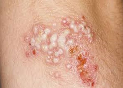
Infected person with HSV-1 has blisters on the mouth
Picture 2: Infected person with HSV-1 has blisters on the mouth.
Image Source :encrypted-tbn0.gstatic.com

Herpes simplex virus- type 2 (HSV-2)

It is a worldwide epidemic. It is sexually transmitted. It is estimated that 417 million people had contracted the infection by 2012. This infection is incurable and lasts a lifetime. Survey shows that most affected are women.

This is because transmission from a man to a woman is easier than from a woman to a man. In 2012 it is estimated that 267 million women and 150 men are infected with HSV-2.

Signs and Symptoms

  • The symptoms are very mild and therefore the infected is unlikely to notice it.
  • Noticeable symptoms are open sores and ulcerations.
  • Fever, body aches and swollen nymph nodes are likely to be felt by the individual.
  • Recurrent ulcerations will occur but usually reduce over time.
  • The infected may experience pain on hips, legs and buttocks before other symptoms.

Transmission

HSV-2 is transmitted when a partner has sexual relations with an infected person especially through fluids in the genitalia, skin and anal area. Infected persons are more susceptible to contracting HIV infection.

Infected persons with HSV-2 living with HIV infections have more recurrent symptoms that infected people with only HSV-2.

Psychosocial Impact

People living with HSV-2 have stress and experience social stigma. The sexual life is affected negatively.

Treatment

Since there is no cure for viral diseases, antiviral medication is provided to the patient in order to reduce the impact and severity of the symptoms. Such medication include acyclovir,famciclovir, valacyclovir.

Prevention

  • Infected individuals should abstain from sexual activities.
  • Steady and correct use of condom will minimize the risk of one getting infected.
  • Pregnant women who have contracted the infection are advised to visit a doctor in order to help them with the situation.


HSV-2 on genitalia
Picture 3: HSV-2 on genitalia
Image Source: encrypted-tbn1.gstatic.com

References :

  1. Clark DC (2003). “Common acute hand infections”. Am Fam Physician 68 (11): 2167–76. PMID 14677662
  2. https://www.nlm.nih.gov/medlineplus/herpessimplex.html
  3. Berger JR, Houff S. Neurological complications of herpes simplex virus type 2 infection. Arch Neurol. May 2008; 65(5):596-600.
  4. Harvey Simon, MD, Editor-in-Chief, Associate Professor of Medicine, Harvard Medical School; Physician, Massachusetts General Hospital. Also reviewed by David Zieve, MD, MHA, Medical Director, A.D.A.M., Inc.
  5. Lewis MA (2004). “Herpes simplex virus: an occupational hazard in dentistry”. Int Dent J 54 (2): 103–11. doi:10.2956/indj.2004.54.2.103. PMID 15119801.
  6. Avitzur Y, Amir J (2002). “Herpetic whitlow infection in a general pediatrician–an occupational hazard”. Infection 30 (4): 234–6. doi:10.1007/s15010-002-2155-5. PMID 12236568.
  7. Herpetic whitlow an occupational hazard, AANA J. 1990 Feb;58(1):8–13. Klotz R.W
  8. Freedberg, Irwin M., ed. Fitzpatrick’s Dermatology in General Medicine. 6th ed, pp.1317, 2063, 2068. New York: McGraw-Hill, 2003.
  9. Bolognia, Jean L., ed. Dermatology, pp.1237-1238. New York: Mosby, 2003
  10. http://infectionnet.org/notes/herpes-viruses/

No responses yet

Lump Behind Ear

Mar 06 2017 Published by under Diseases and Conditions

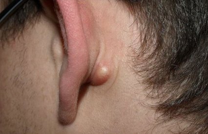
Most lumps are harmless and painless. They are signals for medication. Lumps can either be small or large. Mostly, they are small knots or lumps at the back of the head and just below the ear can be described as a pea-sized. [1, 2]

LUMP BEHIND EAR

Picture 1: A swollen pea lump just behind the ear.
Image Source: www.treatcurefast.com

A lump in the neck or throat is most likely to be one of the following:

Swollen glands– This is usually a sign of infection, such as cold. It may also be accompanied by fever and fatigue. This indicates that there is an infection. When the infection is treated, the fever disappears.

A Cyst – This is a harmless fluid-filled lump that may disappear on its own without treatment. It will feel like a pea and roll under the skin when you press it.

Benign tumor – This are tumors that appear as painless and soft. They are movable lumps that may grow from the salivary gland tissues up to the areas behind the ears. These tumors can either grow or not grow.

Cancer -Harmful cancerous growths can develop behind the ears

CAUSES Of LUMPS BEHIND THE EAR

The following could be the causes of lumps behind the ear.

Abscess

An abscess develops when tissue or cells in an area of the body become infected. The body responds by trying to fight off the bacteria or virus. This happens when the body sends white blood cells to the infected area.

These white blood cells begin accumulating in the damaged location. When large amounts of white blood cells accumulate in the infected area, pus is formed. Pus is a thick, fluid-like product that develops from dead white bloods cells; tissue, bacteria, and other invading substances. They are mostly painful and very warm. [1, 2]

Picture 2 shows Abscess containing pus
Image Source:www.reddit.com

Acne Vulgaris

When the hair follicles on the skin become clogged, acne is formed. Acne can be formed by accumulation of dead cells and oil.

These results to what we call pimples or bumps. They can be formed anywhere. They are mostly found on the face and also behind the ear. These bumps can grow to be large and they are usually painful. [1, 2, 4]

Picture 3 : shows a swollen area is as a result of accumulated cells.
Image Source:mys.yoursearch.me

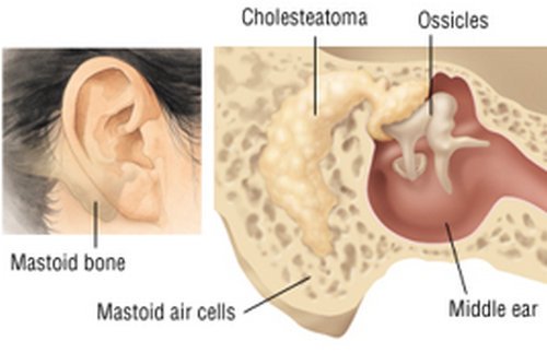
Mastoiditis

When lumps behind the ear overstay without treatment, they may develop serious ear infection called Mastoiditis. This name was developed because the infection is found on the bony protrusion of the ear called the mastoid. This lump may have pus in it and is painful. [2, 6]

Picture

Mastoid bone
The first picture shows an infection caused by an overstayed lump that was untreated. The second picture shows the exact location of the mastoid bone and where the infection occurs.
Image source: www.drugs.com

Lipoma

This is caused when a fatty lump forms between the layers of the skin. Lipoma can form in any part of the body. It is almost completely harmless. Often, it is painless. Mostly they are not detectable on the skin surface but they can grow larger and be felt on the skin surface. [2, 3]

Picture 5: Lipoma which is fairly visible and appears to be harmless.
Image Source: en.wikipedia.org

Dermatitis

Dermatitis is brought about when there is accumulation of dead cells behind the ear. This can also be as a result of cells that are flaking.

Most times it is characterized by lump that is red in color and painful. Stress and differences in climate can cause dermatitis. People with low immune systems such as AIDS are prone to dermatitis. [2, 5]

Picture 6: The infected area is the greyish inflammation on the outline of the back side of the ear.
Image Source:www.skinsight.com

Cancer

Malignant cancer growth such salivary gland and skin cancers could develop behind ears. Lumps from these malignant growths painless and will keep growing over time. [1, 3]

Picture 6: A cancerous growth just behind the ear.
Image Source: cancer.ucsd.edu

How to identify Lumps behind the ear

A person can identify a lump behind the ear by conducting a complete self check on the body. This is effective by the feel of the hand. In children they may resemble chicken pox or pimples.

A regular checkup can be done after ear piercing. Some may be harmless such as lipoma but others can be painful, in that case, being a sign of infection. [7]

Treatment

Different types of lumps of the ear have different treatment techniques. The following are treatment for the various causes of lumps behind the ear.

   1.  Abscess

For some small abscess they may drain and heal without the need of treatment. Applications of heat in the form of a warm funnel can speedup up the healing process. For persistent abscess the doctor can prescribe antibiotics such as broad-spectrum.

If your skin abscess needs draining, you will probably have a small operation carried out under anaesthetic where you remain awake and the area around the abscess is numbed.

During the procedure, the surgeon will make a cut (incision) in the abscess, to allow the pus to drain out. They may also take a sample of pus for testing.

In other cases the pus can also be drained by the use of a needle. [1, 2]

   2.  Acne Vulgaris

  • This is treatable through oral antibiotics for adults.
  • Oral antibiotics are applicable to all patients from mild to severe inflammations with the exceptions of children below eight years of age.
  • For females who are within puberty period, oral antibiotics can be administered together with hormonal therapy for quick recovery.[1, 2, 4]

  3.  Mastoiditis

  • Prescriptions such as oral antibiotics can be administered to a patient.
  • Ear drops can also be an effective way to drain pus.
  • A mixture of olive oil and garlic oil can also be an effective way to cure mastoiditis.[2, 6]

  4.   Lipoma

  • A lipoma that is left alone usually doesn’t cause any problems. However, a dermatologist can treat the lump if it’s uncomfortable.
  • The most common way to treat a lipoma is to remove it through surgery. This is especially helpful for large skin especially if the tumor is still growing.
  • Another option is liposuction where a needle attached to a large syringe, and the area is usually numbed before the procedure.
  • Steroid injections may also be used right on the affected area. This treatment can shrink the lipoma, but it doesn’t completely remove it. [2, 3]

  5.   Dermatitis

  • Topical creams and lotions with corticosteroids like hydrocortisone and betamethasone will help reduce the itching sensation and discomfort.
  • Antifungal agents like ciclopirox and ketoconazole can be prescribed where there is a yeast infection.
  • Cover the area using bandages and dressings so as to totally stop the scratching.
  • Take a soothing bath using uncooked oatmeal or baking soda. [2, 5]

Cancer

Treatment of lumps that are cancerous depends on the stage that they are in. they can be treated or managed either by chemotherapy, radiation or excision. [1, 3]

Precaution

  • When you detect a lump behind your ear, one should pay attention on how long it is going to last.
  • If it is still persistent and maybe it is painless, it is advisable to let your doctor know about the situation.

References :

  1. Neville BW, Damm DD, Allen CA, Bouquot JE. (2002). Oral & maxillofacial pathology (2nd ed.). Philadelphia: W.B. Saunders. ISBN 0721690033.
  2. Bancroft LW, Kransdorf MJ, Peterson JJ, O’Connor MI (October 2006). “Benign fatty tumors: classification, clinical course, imaging appearance, and treatment”. Skeletal Radiol. 35 (10): 719–33. doi:10.1007/s00256-006-0189-y. PMID 16927086.
  3. James, William D.; Berger, Timothy G.; Elston, Dirk M. (2005). Andrews’ Diseases of the Skin: Clinical Dermatology (10th ed.). London: Elsevier. ISBN 0-7216-2921-0.
  4. Tuchayi, Sara M.; Makrantonaki, Evgenia; Ganceviciene, Ruta; Dessinioti, Clio; Feldman, Steven R.; Zouboulis, Christos C. (17 September 2015). “Acne vulgaris”. Nature Reviews Disease Primers.
  5. Bershad, SV (1 November 2011). “In the clinic. Atopic dermatitis (eczema)”. Annals of internal medicine 155
  6. MedlinePlus Medical Encyclopedia. Retrieved July 30, 2003.
  7. O’Handley JG, Tobin EJ, Shah AR. Otorhinolaryngology. In: Rakel RE, ed. Textbook of Family Medicine. 8th ed. Philadelphia, PA: Saunders Elsevier; 2011:chap 19.
  8. http://www.google.com
  9. http:// www.healthline.com
  10. www.just-health.net. Last Updated 15 January, 2016

No responses yet

Pitted Keratolysis

Mar 05 2017 Published by under Diseases and Conditions

Background

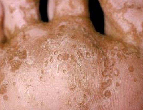
  • Pitted keratolysis refers superficial skin condition that affects the feet. The feet are covered by pits that are largely asymptomatic.
  • In many cases, pitted keratolysis exhibits random episodes of occurrence and disappear just as spontaneously [1]. As such, the condition can affect the person for several years if it is left untreated.
  • To effectively treat the condition the wet and warm conditions that promote the reproduction and growth of the causative bacteria have to be controlled first.
  • Most prevalent among individuals with hyperhidrosis (profuse sweating) especially when they have to go long periods of time in closed shoes [2].

Symptoms

  • Pits – 1  to 3 mm in diameter [3]
  • However, the pits may overlap leading to a larger lesion [4].
  • Smelly feet. This is the major cause of anxiety among the sufferers
  • The heel, the forefoot, or both become covered with punched out sores that make them white in color. This is because these areas are subjected to the weight of the body [2].
  • In a certain type of the condition, some red spots appear on the sores [2, 4]
  • Sometimes the hands and fingers are also affected [5]
  • The sores are usually asymptomatic and most people seek medical attention due to the foul smell and embarrassment caused by the sores [1, 3]
  • Some patients claim to feel some irritation but it is mild.
  • The skin in some areas of the feet may be white and swollen.


characteristic of pitted keratolysis
Image 1: Lesions characteristic of pitted keratolysis
Picture Source: skinsight.com

Causes

  • The condition is caused by a host of gram-positive bacteria. They include: Kytococcus sedentarius, Dermatophilus congolensis, streptomyces and actinomyces [5].
  • Enclosed feet, hyperhidrosis and high skin pH offer the optimum conditions for these bacteria to reproduce and produce digestive enzymes-proteases [3].
  • The proteases digest the foot skin (stratum corneum) proteins resulting to the cater-like pits which develop into sores [5]
  • The foul odor has been attributed to the sulphur compounds that the bacteria release as products of metabolism [6]

 profuse hyperhidrosis
Image 2: Hands of a person with profuse hyperhidrosis
Photo Source: homeremediesorg.com

Who is at Risk?

  • Pitted keratolysis has been observed to be more likely to infect men. This may be because men are more inclined to wear occlusive, non-breathing footwear to work or school. [6]
  • People of all ages can be infected by the bacteria and subsequently get the illness.
  • It is a common condition among athletes, construction and industrial workers, and people in the military because of the requirements that they be in boots most of the time [7]
  • Anyone working in wet/moist conditions such as people working in swimming pools, spas, and paddles [6,7]
  • Sweaty feet or palms also predispose individuals to the illness [2, 8].

Diagnosis

  • Clinical assessment of the patient’s signs and symptoms.
  • Using as swab to take a probe for lab analysis to help identification of the causative microorganism.
  • In case there is suspicion that the infection has been caused by different strains of fungi, the skin can be scrapped for examination and confirmation [4].
  • Skin biopsies are rarely necessary since the diagnosis can be done easily owing to the characteristic pits and the odor [9].
  • The Wood’s ultraviolet light test is inconsistently helpful. The affected surface shows a distinctive red fluorescence [9].
  • The causative bacteria may be collected from the pits. They can then be cultured on a medium with brain-heart infusion agar. The culture is placed under nitrogen and carbon dioxide. Temperature should be 98.6oF (37o).

Management

Preventive measures

  • Minimize the use of closed footwear and where it is unavoidable wear shoes that fit properly to limit friction on the [6].
  • Keep feet always dry. After washing them, one can use a hair blow dryer to dry them up [4].
  • Wear clean cotton socks. They should be washed and changed frequently [pcds].
  • Those prone to the illness should also avoid wearing a pair of shoes for more than two consecutive days [4].
  • Use innersoles that are designed to absorb moisture when wearing closed shoes. Just like socks, the innersoles should be changed frequently.
  • Whenever possible, the feet should be let out of the shoes to breathe [5, 7].
  • Don’t share towels with others [10]
  • Use an antiseptic to wash the feet twice a day. Dry them properly [10]
  • People involved in sports and working out should not linger in their footwear after sweaty exercises. After the exercise or workout, it is recommended that one wears open shoes with adequate ventilation.

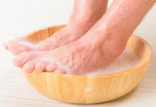
Feet require special treatment to manage Pitted Keratolysis
Image 2: Feet require special treatment to manage Pitted Keratolysis
Picture Source: wisegeek.com

Cure and Home Treatment

  • Palmoplantar hyperhidrosis causes profuse sweating of the palms and feet. It is necessary to treat the condition first before trying to treat pitted keratolysis.
  • A solution of 5% formaldehyde solution is effective in cases of extremely wet feet where other techniques used to dry the feet have not succeeded [1]. The solution can only be obtained with a prescription and it is relatively expensive.
  • Prescribed strength-antiperspirant powder applied in the socks and shoes will minimize the perspiration. As a result the environment will be unsuitable for the bacteria to thrive [10].
  • When used in combination with the preventive measures, topical antibiotics work in a span of 3-4 weeks [6]
  • Clindamycin, erythromycin preparations or Fucidin cream applied twice a day has also been noted t treat pitted keratolysis [2]
  • Many cases have been reported where individuals have successfully treated the condition by applying acne medication with 10% benzoyl peroxide the part of foot that is infected [1].
  • Oral erythromycin has also been reported to be effective in some instances [2, 6].
  • In severe cases injections of the botulinum toxin may be administered [11].
    Is Pitted Keratolysis contagious?
  • Unlike most of other infections caused by bacteria, pitted keratolysis is not contagious a contagious medical condition [4].
  • However, it is advisable to maintain good personal hygiene. It is also recommended that people do not share personal objects of hygiene such as towels amongst each other because it may lead to secondary infections [4].

References :

  1. Pitted Keratolysis. Treatment [Internet]. 2012 [cited 20 January 2016]. Available from: http://www.pittedkeratolysis.com/treatment/
  2. Dermnetnz.org. Pitted keratolysis. DermNet NZ [Internet]. 2016 [cited 20 January 2016]. Available from: http://www.dermnetnz.org/bacterial/pitted-keratolysis.html
  3. Podiatrytoday.com. A Review Of Treatment Options For Pitted Keratolysis | Podiatry Today [Internet]. 2016 [cited 20 January 2016]. Available from: http://www.podiatrytoday.com/blogged/review-treatment-options-pitted-keratolysis
  4. Medicalpoint.org. Pitted Keratolysis – Treatment, Pictures, Symptoms, Contagious [Internet]. 2016 [cited 20 January 2016]. Available from: http://medicalpoint.org/pitted-keratolysis/
  5. Emedicine.medscape.com. Pitted Keratolysis: Background, Pathophysiology, Epidemiology [Internet]. 2016 [cited 20 January 2016]. Available from: http://emedicine.medscape.com/article/1053078-overview#showall
  6. Pcds.org.uk. Pitted keratolysis [Internet]. 2016 [cited 20 January 2016]. Available from: http://www.pcds.org.uk/clinical-guidance/pitted-keratolysis
  7. Gustrength.com. Pitted and Smelly Feet: Pitted Keratolysis – Ground Up Strength [Internet]. 2016 [cited 20 January 2016]. Available from: http://www.gustrength.com/health:pitted-and-smelly-feet-pitted-keratolysis
  8. Khachemoune A, T. Klausner B. Smelly, Macerated Feet Diagnosis: Pitted keratolysis (PK) | The Dermatologist [Internet]. The-dermatologist.com. 2016 [cited 20 January 2016]. Available from: http://www.the-dermatologist.com/article/851
  9. Pitted keratolysis. Indian Journal of Dermatology, Venereology and Leprology [Internet]. 2005 [cited 20 January 2016];71(3):213-215. Available from: http://www.ijdvl.com/article.asp?issn=0378-6323;year=2005;volume=71;issue=3;spage=213;epage=215;aulast=
  10. Med-health.net. Get Rid of Pitted Keratolysis Completely | Med-Health.net [Internet]. 2016 [cited 20 January 2016]. Available from: http://www.med-health.net/Pitted-Keratolysis.html
  11. Skinsight.com. Pitted Keratolysis in Adults: Condition, Treatment and Pictures – Overview | skinsight [Internet]. 2016 [cited 20 January 2016]. Available from: http://www.skinsight.com/adult/pittedKeratolysis.htm

No responses yet

Immature granulocytes

Mar 04 2017 Published by under Diseases and Conditions

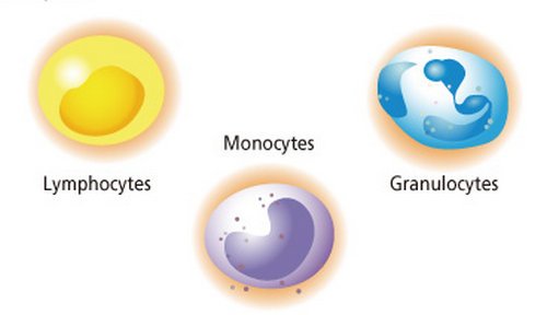
Granulocytes are the white blood cells that have small granules that contain proteins. The specific granulocytes are: neutrophilis, eosinophils, and basophilis [1,2].

  • Granulocytes come from stem cells in the bone marrow of a person. The process of differentiation of these stems from the pluripotent hematopoietic stem cell to granulocytes is called granulopoiesis.
  • Immature granulocytes are part of the immune system which responds to infection or inflammation. Immature granulocytes are an indication of an early stage of an infection.

Leucocytes

Types of leucocytes

What are immature granulocytes

  • Immature granulocytes are white blood cells which only appear when the bone marrow is activated to fight of an infection they not only circulate blood but even if a healthy individual has a small count there should be no cause off concern when immature granulocytes. When absolute number of immature granulocyte increase significantly, this is a sign of infection [3,5,6].
  • The neutrophil portion of the blood count usually goes upon when the immature granulocytes high levels begin to appear. This is a clue that there is something going on in the body such as bacterial infection, inflammatory disease, trauma in the body, use of steroids or maybe cancerous conditions.
  • The elderly and young infants experience increase in immature granulocytes without neutrophils increase. [1]
  • In the event this happens the increase needs to be thoroughly investigated.

Immature granulocyte count

  • Apart from blood from neonates or pregnant women immature granulocytes appearance in the peripheral blood indicates an early stage response of an infection and inflammation or other stimuli of the bone marrow [4].
  • Areas of research on the diagnostic importance of circulating immature granulocytes are focused on early, rapid discrimination of bacteria from infections that are viral particularly in the young children hence recognizing bacterial infection in neonates, and early bacterial recognition.[2, 3]
  • For better understanding of how to get accurate immature granulocyte count we need to understand the differential blood count
  • Differential gives relevant percentage of each type of white blood cells and also helps to reveal abnormal white blood cell population.
  • Differential blood count could be performed using the following two methods.
  • Automated differential blood counts: Automated hematology instruments that use multiple methods and parameters used to count and identify five major types of white blood cells in blood.
  • Manual differential blood count: Manual count is done by visual examination of sample peripheral blood smear.
  • Automated differential blood count consumes less time and costs less than routine blood smear examination. The automated technique allows thousands of the white blood cells to be examined. Visual examination can only allow 100-200 white blood cells to be examined. [4]

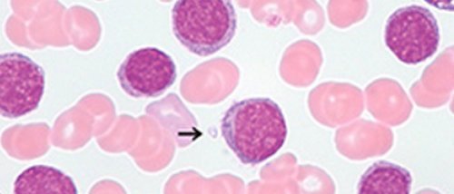
Cell images

                    Cell images

Propose of preferential blood count

  • Blood differential test measures the percentage of each white blood cell that individuals have in the blood. It also determines if there are any abnormalities or immature cells

Normal range

  • The normal range describes where 95% of most health people population will lie.[5]
  • It is very useful for biomedical scientists and haematology clinicians to have appropriate clinical details added to the request so that interpretation and best clinical advice can be given on the report where appropriate [7].
  • A person diagnosed with CML should have routine blood tests which include complete blood count (CBC), blood differential and liver functioning tests which are done on a regular basis so as to help monitor treatment response.
  • A complete blood count (CBC) which measures the number of white blood cells and platelets in a patient’s blood sample should be routinely monitored in CML patients.[6, 7]

Neutrophils

  • These are a type of white blood cells involved in fighting infection. It’s of importance that they remain at adequate levels. As with platelets, neutrophils levels may be depressed in patients on myelosuppressive therapy such as imanitibbmesylate the normal range of the percentage of neutrophils is between 45-70% [9]
  • More important than the neutrophils percentage is the absolute neutrophil count. Which will fall between 1.0-8.0 k/ul.
  • The reason the absolute neutrophil count (ANC) represents the clear clinical picture better than the percentage of neutrophils is that, in cases where blood counts are suppressed by the therapy, percentage of neutrophils are higher when overall counts are law. One can calculate the ANC by multiplying the percentage of neutrophils by the percentage of bands by the total number of white blood cells, the number of white bands is usually quite low or zero.[8, 9]

Basophils

  • The basophils should remain within the normal range generally between zero-two percent.
  • Some medics believe that the absolute basophil count is of more importance than the basophils percentage and should fall between 0-0.3k/ul.

When low counts are of concern

  • This depends on some individuals, the larger clinical picture and the therapy receive. In general for patients on imatianib mesylate therapy, a disease in those may be warranted by the following levels: the WBC less than 1.0k/ul; platelets less than 50k/ul;haemoglobin less than 10.0gm/Dl;and ANC less than 1.0k/ul.

Performance of blood differentials and CBCs

  • All patients taking IM should have their blood counts monitored closely. Complete CBCs should be monitored closely on weekly intervals in chronic phase patients during first the first month of IM treatment.
  • If counts are stable monitoring may occur monthly or even longer if appropriate. Patients in accelerating or blast crisis should have CBCs performed more often

Interpretation of full blood count

  • A full blood count should be interpreted with reference to the clinical picture and other pathology results. Cautions must be given to certain abnormal results such as low platelets that may be artefact and rapids should be considered the laboratory will usually be investigate the abnormalities that may be haemotological in nature may accessing the blood film, the event of serious abnormalities that require agent attention such as acute leukemia, and severe hemolysis, the laboratory staff will alert the on-call haemotolic doctors.

Patients’ characteristics

  • Patients whose age ranged between 19-88 years and their median ICU length of stay ranged 3-64 days were enrolled in a study to characterize disease severity the SAPs score was determined on the day of ICU admission the population had a median score of 37 showing expected ICU mortality rate of 19%.[10]

References:

  1. Sysmex-europe.com. Immature Granulocyte (IG) count – Sysmex Europe GmbH [Internet]. 2016 [cited 16 January 2016]. Available from: http://www.sysmex-europe.com/academy/knowledge-centre/sysmex-parameters/immature-granulocyte-ig-count.html
  2. Enkivillage.com. When Is Immature Granulocytes Too High and What Causes It? – EnkiVillage [Internet]. 2016 [cited 16 January 2016]. Available from: http://www.enkivillage.com/immature-granulocytes.html
  3. Healthline. Granulocytosis [Internet]. 2016 [cited 16 January 2016]. Available from: http://www.healthline.com/health/granulocytosis
  4. Nierhaus A, Klatte S, Linssen J, Eismann N, Wichmann D, Hedke J et al. Revisiting the white blood cell count: immature granulocytes count as a diagnostic marker to discriminate between SIRS and sepsis – a prospective, observational study. BMC Immunol. 2013;14(1):8.
  5. Clinlabnavigator.com. Immature granulocytes replaced bands as an indicator of infection | ClinLab Navigator [Internet]. 2016 [cited 16 January 2016]. Available from: http://www.clinlabnavigator.com/immature-granulocytes.html
  6. pdrhealth. Leukemia Diagnosis – Diseases and Conditions – PDR Health [Internet]. 2016 [cited 16 January 2016]. Available from: http://www.pdrhealth.com/diseases/leukemia/diagnosis
  7. Che Y, Shen D, Zhang S, Qi J. Identification of immature granulocytes in cancer chemotherapy patients by cell counting vs. microscopic examination of blood smears. Molecular and Clinical Oncology [Internet]. 2014 [cited 16 January 2016];2(2):207-211. Available from: http://www.spandidos-publications.com/mco/2/2/207
  8. Emedicine.medscape.com. Differential Blood Count: Reference Range, Interpretation, Collection and Panels [Internet]. 2016 [cited 16 January 2016]. Available from: http://emedicine.medscape.com/article/2085133-overview#showall
  9. Senthilnayagam B, Kumar T, Sukumaran J, M. J, Rao K. R. Automated Measurement of Immature Granulocytes: Performance Characteristics and Utility in Routine Clinical Practice. Pathology Research International. 2012;2012:1-6.
  10. HealthTap. Top 11 Doctor Insights on what does high immature granulocytes mean – HealthTap [Internet]. 2016 [cited 16 January 2016]. Available from: https://www.healthtap.com/topics/what-does-high-immature-granulocytes-mean


No responses yet

Bruised Sternum

Mar 03 2017 Published by under Diseases and Conditions

The Sternum

  • Also known as the breastbone, the sternum is a long, T-shaped flat bone found at the center of the human thorax [1].
  • At its upper end, the sternum connects to the clavicles.  On both sides, it is articulated to ribs [2].
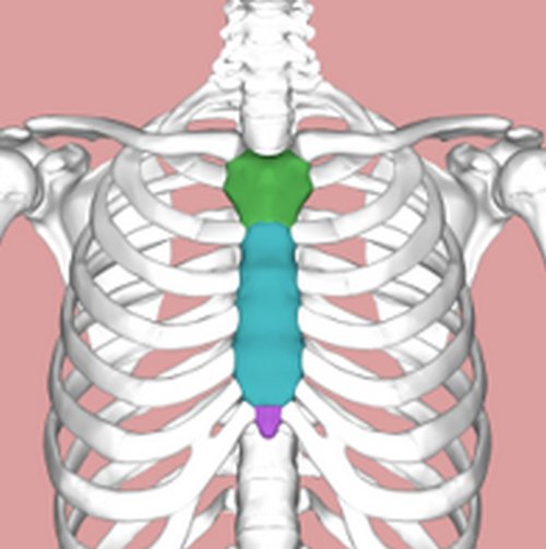
  • It is made up of three parts i.e. the xiphoid process, the manubrium, and the body (also known as gladiolus) [3].

    : An illustration of the sternum

Figure 1: An illustration of the sternum: green part (manubrium);
Blue part (body); purple part (xiphoid process)
Source: Wikipedia.org

  • Its T-shape resembles a sword that is pointing downward. The shape inspired the names of its parts: gladiolus means “sword” in Greek, manubrium handle, and xiphoid means sword-shaped [4].
  • Together with the rest of the thoracic wall, the sternum’s function is to protect the inner, more delicate organs such as the lungs, heart, the esophagus, and major blood vessels against physical injury [1, 2].

Sternum Injury

  • Sternum injury (sternum contusion) happens as a result of direct physical impact on the breastbone [5].
  • It is a rare type of injury and usually occurs in car accidents and rarely in sports involving collisions such as rugby and those that entail use of hard equipment such as balls or rackets [5].
  • Due to the involvement of the chest in most physical exercises, a bruised sternum hampers working out and participation in sports.

Causes of Bruised Sternum

  • In car accidents, if one is wearing the seatbelt it may be the cause of the injury as seat belt tries to stop driver or passenger [2].
  • If no seat belt is worn, the impact as the driver slams on the steering may cause an impact that harms not only the sternum but other thoracic organs [2].


Image 2: An illustration of the impact of a steering wheel in an accident
Source: worldroadsafety.org

  • Contact sports.
  • Whooping cough:  A whooping cough may be too violent that it causes injury to the sternum in young children [2].

Symptoms : What does a bruised sternum feel like

  • Pain on the sternum due to the physical impact
  • Pain while breathing, coughing, bending and sneezing
  • Tenderness on the front part of the chest lying over the sternum
  • Localized swelling on the center of chest just over the sternum
  • Bruises may appear on the chest later on


showing visible bruises on the chest

Picture 3: A picture showing visible bruises on the chest

Image Source: treatcurefast.com

Treating Bruised Sternum

  • Sternum injury is usually not severe and should heal in a matter of weeks.
  • However, if the pain is severe medical attention should be sought. The doctor may take x-rays to confirm that no fracture was suffered on the ribs or sternum [5].
  • Remedies include:

Anti-inflammatory medication

These are meant to be taken under prescription. They are designed to address any inflammation caused by the impact as well as the side-effects of the inflammation. The effects attributable to inflammation may include: Swelling, localized pain, change in color (to redness) and an increase in temperature (fever) [2].

Icing

It serves two main purposes: relieving the pain and minimizing the inflammation-related swelling [6].
The ice can either e rubbed along the point of injury.
Alternatively, it can be applied along the entire thoracic region (will require more ice)
The ice should be applied 3 or 4 times per day, with each rub lasting up to 10 minutes [2].
Direct contact with the skin may be harmful. As such, the ice should be wrapped in a piece of cloth.

Change in lifestyle

To avoid causing further injury to the sternum, it is imperative to adjust activities one is involved in during the period of recovery. First, one has to avoid any form of heavy lifting. In addition, contact sports, intense movements that cause irritation to the chest, and bending over should be avoided [2, 6].
For individuals that smoke, avoiding the cigarettes will reduce the healing time considerably [7].
If there is a cough accompanying the injury, the person should take medication to treat it as well.

Change in diet

The medical officer may suggest a change in diet to promote faster recovery.
The changes will involve taking a diet with nutrients that are crucial to the healing of the bones. A diet rich in calcium is highly recommended [6].

Supplements

Taking in bromelain supplements has been shown to reduce the swelling. It also minimizes the pain, and they are believed to provide essential digestive enzymes. Eating pineapples has a similar effect [7].
Vitamin C and K food supplements also help in the healing process. The two are essential co-factors in some bio-physiological processes, and they have been observed to reduce the recovery period significantly [2, 7].

Micro-current therapy

This is yet another way to treat bruised bones and sternum in particular.
It is recommended for individuals that have to resume their normal schedules fast since it quickens recovery process [8].
The area of the chest affected by the bruise is exposed to of electric current. The current has to be quite low due to the sensitivity and tenderness of the tissues around the point of injury [7, 8].
The electric current is aimed at correcting the electrical fields of the body that were disrupted because of the trauma resulting from the cause of injury [7]. As such, the method serves to heal not only the bones but the surrounding tissues as well.

Recovery Period

  • The recovery period for the bruises ranges between 2 and 4 weeks.
  • Patience and rest are crucial during this period [9].
  • During the recovery period one should avoid physical activities to avoid worsening the injuries.
  • Once recovered, one should start with the light-impact physical activities, and this should be under the guidance of a doctor [7]. The light impact training includes swimming and biking to re-acclimatize the body.

Complications

  • Due to the lack of severity, there are no complications attributed to bruised sternums. However, if the patient does not take proper caution during the treatment period, the injury may become severe.
  • Severe pain may be a sign of a fractured sternum and/or ribs. X-rays are necessary in such situations to confirm whether it is a fracture or bruise [9].

Caution

  • Although seat belts have been noted to cause sternum injuries, they prevent injuries that may prove to be worse, including to internal organs such as the neck, spinal cord, and the brain. Therefore, wearing seatbelts is recommended as a method of ensuring driver and passenger safety on the road [10].

References:

  1. Teachmeanatomy.info. The Sternum – Body – Manubrium – Xiphoid – TeachMeAnatomy [Internet]. 2016 [cited 16 January 2016]. Available from: http://teachmeanatomy.info/thorax/bones/sternum/
  2. Helpyourback.org. 4 At-Home Remedies For Bruised Sternum Treatment [Internet]. 2014 [cited 16 January 2016]. Available from: http://helpyourback.org/health/at-home-remedies-for-bruised-sternum-treatment
  3. Wikipedia. Sternum [Internet]. 2016 [cited 16 January 2016]. Available from: https://en.wikipedia.org/wiki/Sternum
  4. InnerBody. Sternum [Internet]. 2016 [cited 16 January 2016]. Available from: http://www.innerbody.com/image_chest1/skel16.html
  5. Sportsinjuryclinic.net. Bruised Sternum [Internet]. 2016 [cited 16 January 2016]. Available from: http://www.sportsinjuryclinic.net/sport-injuries/chest-abdomen-pain/bruised-sternum
  6. Pocock C. Bruised Sternum | Med Health Daily [Internet]. Medhealthdaily.com. 2016 [cited 16 January 2016]. Available from: http://www.medhealthdaily.com/bruised-sternum
  7. Treat, Cure Fast. Bruised Sternum Symptoms, Pain, Treatment and How to Heal a Bruised Sternum [Internet]. 2014 [cited 16 January 2016]. Available from: http://www.treatcurefast.com/bruises/heal-bruises/bruised-sternum-symptoms-treatment-pain-heal
  8. Orthopedics.ygoy.com. How to Treat a Bone Bruise? – Orthopaedics [Internet]. 2016 [cited 16 January 2016]. Available from: http://orthopedics.ygoy.com/2011/09/06/how-to-treat-a-bone-bruise
  9. Treat, Cure Fast. Bruised Sternum Symptoms, Pain, Treatment and How to Heal a Bruised Sternum [Internet]. 2014 [cited 16 January 2016]. Available from: http://www.treatcurefast.com/bruises/heal-bruises/bruised-sternum-symptoms-treatment-pain-heal
  10. Worldroadsafety.org. World Road Safety Partnership [Internet]. 2016 [cited 16 January 2016]. Available from: http://www.worldroadsafety.org/php/allSafetyPgms.php?cid=8&pid=59

No responses yet

Warts on Tongue

Mar 01 2017 Published by under Diseases and Conditions

Have you ever tried to eat something and the mouth is very sore such that anything that goes into the mouth comes out as fast? You admire tasty food only with your eyes because even a little salt irritates your mouth.

Was it a sore that caused this particularly on the tongue? Well if it was, though rare, this probably was not a usual sore. It could have been a wart. Yes, a wart on the tongue.

What is a Wart on Tongue?

A wart is also known as Verruca Vulgaris (1,2). It is a non-cancerous lesion of the skin and also affects the mucous membrane (2, 3, 4). It can affect any part of the body from the skin, genitals, toes and the tongue is one of those areas.

It is however reported in literature that occurrence on the tongue is quite uncommon (2).Of the few it effects on the tongue, majority are immunocompromised for example from HIV infection. (1)

Wart on tongue
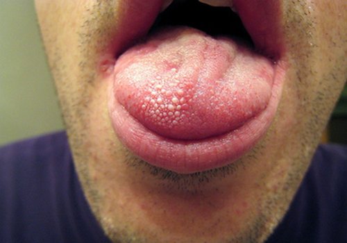
Image 1 : Wart on tongue
Picture Source : medicalpoint.org

How does one get wart on tongue and is it infectious?

Wart on tongue is infectious (1, 6) and is usually transmitted when one gets into contact with another person with a wart. This contact may be through kissing or oral sex. Indeed, many cases reported have been linked to oral sex practice (6). In oral sex, the person will get from an individual with this type of wart in the genitals.

What are the signs and symptoms of wart on tongue?

The signs and symptoms commonly reported are (1, 6, 7);

  • The person will have cauliflower like sores on the tongue. They appear as raised bumps on the skin (8)
  • The sores are rough in texture
  • The color may be white or resemble the color of the mouth
  • Pain on the tongue and difficult moving it
  • The sores could be single or they could be multiple sores
  • There will be difficult eating, talking, chewing or even opening the mouth depending on the exact location of the wart
  • Bleeding from the tongue if one tries to rupture the lesion

What causes warts on tongue?

  • The microorganisms attributed to cause the wart on tongue is a virus.
  • This virus is called Human papillomavirus (HPV). It is the same virus that is associated with cervical cancer and other forms of warts in the body, though the genotypes involved in each case are different (2, 4, 9).
  • This virus is transmitted through sexual contact and kissing to cause wart on tongue (9)

What is the treatment for wart on tongue?

Wart on tongue is actually harmless though it causes discomfort. The lesions are typically known to be self-limiting, meaning that they can appear and resolve by themselves without any intervention. However, they may also progress to cause immense discomfort to an individual.

The spontaneous healing depends on the person’s immunity and the type of virus involved (2).

One must seek treatment if the wart is extensive, has been there for more than 2 years, causing discomfort and is spreading (4).

In addition, they may obscure the signs and symptoms for oral cancer and therefore, one must be keen to be checked by a doctor to rule out malignancy lesions (10).

Since this is a viral infection, it poses a challenge to treat. However, some medications and remedies can be used to provide comfort and heal the lesions. However scaring may occur after the treatment.

Treatment must be sought form a qualified medical practitioner. Excision or rupturing of the wart at home or by an unqualified personnel should be discouraged as it may cause bleeding, spread of the virus to other areas and injury to the tongue surface (3, 8)

Treatment for warts on tongue and home remedies

  1. Antiviral medications can be applied on the wart. An example is acyclovir topical application. This is commonly used in patients who are HIV positive (1,4). It must be prescribed.
  2. Salicylic acid topical application will reduce the pain (4). They remove the dead skin surface cells promoting healing of the wart (7)
  3. Several surgical treatments including  cryotherapy using liquid nitrogen , electrocauterization and laser therapy have all been tried(2,4,8)
  4. Pain medication to treat the pain. The doctor may prescribe a topical analgesic to reduce the pain (1) to allow eating
  5. Eat soft foods at a comfortable temperature. Identify those beverages and foods that cause minimal discomfort. Avoid highly spiced foods and citrus juices.

Some home remedies have been identified and include(1,4)

  • Applying a banana peel on the tongue several times a day
  • Rubbing a cut raw potato on the affected area
  • Appling aloe Vera juice using a cotton swab
  • Garlic- crush some garlic and cover the affected area
  • Tea tree oil-tea tree oil has been found to have some antiseptic properties. Apply with a cotton wool ball several times a day

How can one prevent wart on tongue?

To prevent the wart on tongue, avoiding contact with individuals who have the warts is key. The commonest mode of transmission is through oral sex, therefore one should avoid oral sex. If this is unavoidable, condoms must be used. However, this does not guarantee 100%protection (6).

Though it is not required that an infected person discloses this to persons they are likely to get contact with, they are encouraged to be socially responsible in order to avoid transmission (1).

If one has a genital wart, it is important that they seek treatment to avoid transmission to the tongue since the same organism can be transmitted through contamination (8)

If one has a wart, they should avoid rupturing it to prevent further spread to other areas (3, 8)

Conclusion

Warts on tongue is a viral non-cancerous lesion that affects the tongue tissue, common among the immunocompromised persons. It is contagious and can be self-limiting resolving without any treatment. When treatment is required, it must be sought from a qualified practitioner to prevent injury to the tissues, spread to other parts of the body and to rule out oral cancer

Reference :

  1. Medicalpoint.org. Warts on Tongue – Pictures, Symptoms, Causes, Treatment [Internet]. 2016 [cited 19 January 2016]. Available from: http://medicalpoint.org/warts-on-tongue/
  2. Ahmet Ural B. Verruca vulgaris of the tongue: a case report with literature review. Bosnian Journal of Basic Medical Sciences [Internet]. 2014 [cited 19 January 2016];14(3):136. Available from: http://www.ncbi.nlm.nih.gov/pmc/articles/PMC4333997/
  3. WiseGEEK. How Can I Remove Tongue Warts? (with pictures) [Internet]. 2016 [cited 19 January 2016]. Available from: http://www.wisegeekhealth.com/how-can-i-remove-tongue-warts.htm
  4. Emedicine.medscape.com. Nongenital Warts: Background, Pathophysiology, Epidemiology [Internet]. 2016 [cited 19 January 2016]. Available from: http://emedicine.medscape.com/article/1133317-overview
  5. Medicalpoint.org. [Internet]. 2016 [cited 19 January 2016]. Available from: http://medicalpoint.org/wp-content/uploads/2011/11/warts-on-tongue.jpg
  6. Healthcentre.org.uk. Oral Warts Treatment | Removing Warts using Oral Treatments [Internet]. 2016 [cited 19 January 2016]. Available from: http://www.healthcentre.org.uk/cosmetic-treatments/wart-oral-warts-treatment.html
  7. Dermnetnz.org.  Warts, verrucas, human papillomavirus infection. DermNet NZ [Internet]. 2016 [cited 19 January 2016]. Available from: http://www.dermnetnz.org/viral/viral-warts.html
  8. Healthline. Warts [Internet]. 2016 [cited 19 January 2016]. Available from: http://www.healthline.com/health/skin/warts#Overview1
  9. Healthline. Human Papillomavirus Infection [Internet]. 2016 [cited 19 January 2016]. Available from: http://www.healthline.com/health/human-papillomavirus-infection#Overview1
  10. Healthline. What Does Mouth Cancer Look Like? [Internet]. 2016 [cited 19 January 2016]. Available from: http://www.healthline.com/health-slideshow/what-does-mouth-cancer-look#2

No responses yet

© 2023 Healthooze.com. All Rights Reserved. Privacy Policy